BackReturn Home

Cell Overall Organization

Introduction

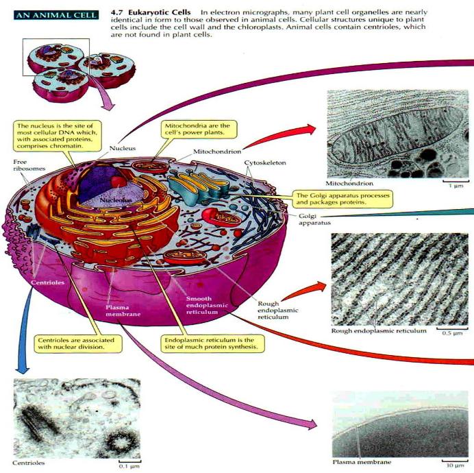
An overview of animal cell structures and what they do will provide an introduction to more detailed articles on how cells work. There are two independent yet related aspects to this. Cells have an intimate triadic relationship with the Organs of the body and with the Host human being. The host Cell in any organ tissue also has an intimate triadic relationship with its internal Organelles and its Enzyme catalyzed chemical reactions. Self-similar structure is pervasive in phenomenal experience. Cells operate in a self-similar way to complex corporations consistent with the structural dynamics of the cosmic order as delineated by System 4. Since the same self-similar patter keeps recurring in different disguises, the analogy with a corporation will help in showing how all cells work.

Cell Overview

Fig. 1

Cell Organelles & Processes

Plasma Membrane

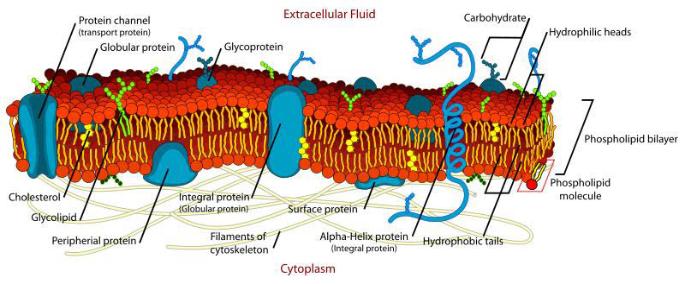
The plasma membrane of the cell is an organelle of primary importance. It consists of a phospholipid bi-layer embedded with thousands of trans-membrane proteins that communicate across from outside to inside and vice versa with related compounds attatched and adjacent. The membrane relates the cell interior to its external market environment. We can compare this to the Marketing domain of a large corporation that relates the cell's capabilities to market needs.

Fig. 2

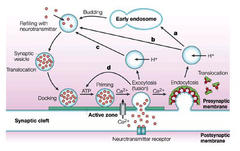
The cell membrane also exports cell products through exocytosis and imports extracellular materials of many kinds through endocytosis. Figure 3 illustrates how exocytosis works cyclically at the synapse of a neuron. Membrane export compares to a shipping department and the import of raw materials through endocytosis compares to a purchasing function. Both are associated with the Production Department of a corporation. The plasma membrane is multifunctional.

Fig. 3

Signaling Cascades

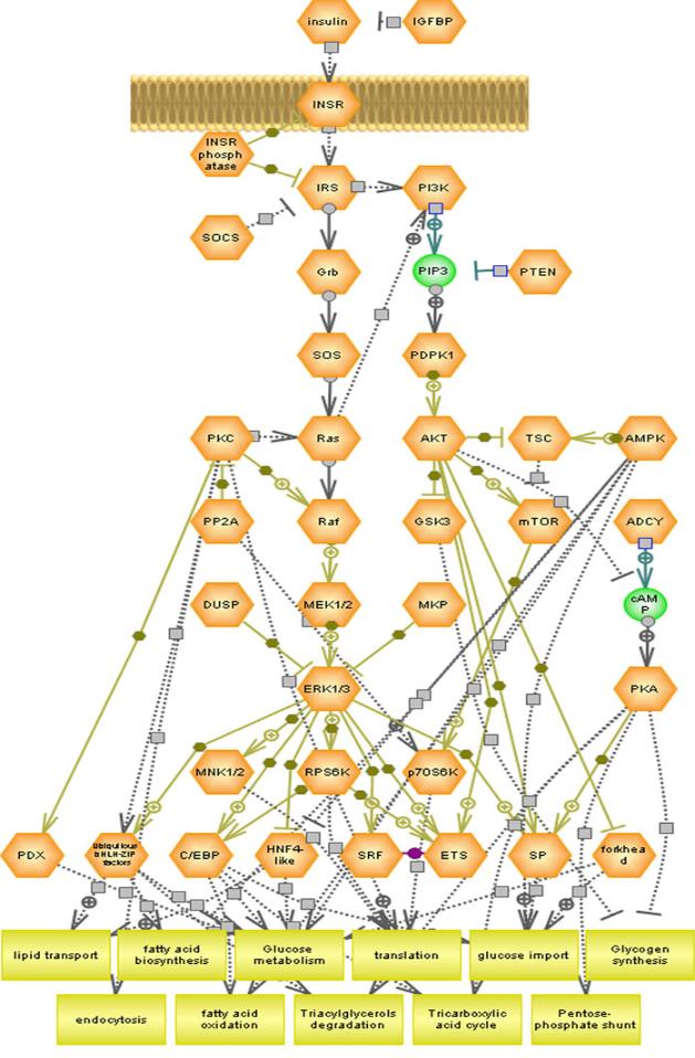
Signaling cascades communicate between organelles in a highly complex maze of interactions. There is huge diversity of signaling pathways and methods possible and a great number of them can operate in parallel often with cross talk between them. The insulin pathways below initiate from a kind of single kind of receptor. There are hundreds of different receptor kinds. A great many of each kind work in parallel to regulate the internal Organization of the cell.

Fig. 4

Cell Nucleus

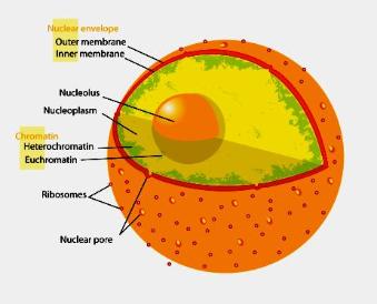
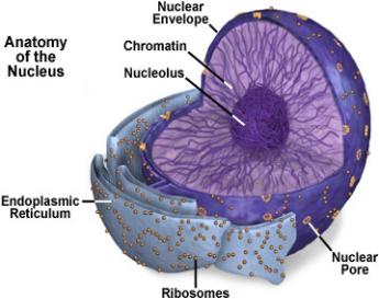
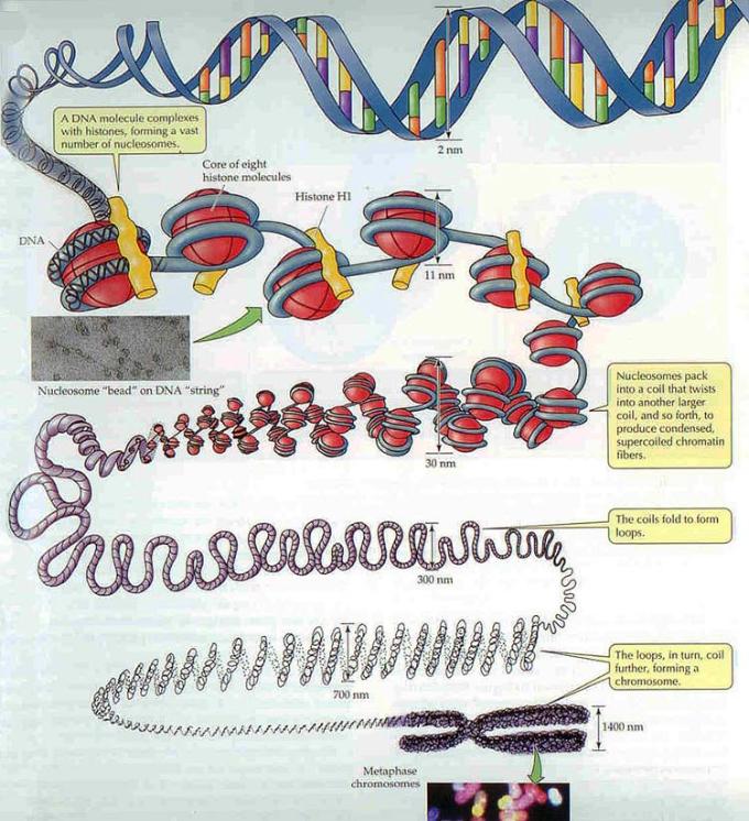
Some signaling to the nucleus is like Sales orders to Engineering to design proteins for export elsewhere in the body. Some are orders to Engineering to make protein and other chemicals for cell maintenance or replication. Most of the latter communication is initiated by Calcium ion signaling. The cell nucleus is enclosed in a double membrane with a few thousand nuclear pores connecting inside and outside. Membrane embedded proteins regulate trans-membrane processes. Inside is the chromatin that has DNA wrapped around histone protein complexes like string around a series of balls that form a very long series of nucleosomes. The chromatin is normally tangled up like spaghetti. If the DNA was strung straight out end to end, it would be nearly two meters long, so it is highly compressed in the nucleus. For cell division, the chromatin replicates, then it goes through a series of additional folding to form chromosomes. The outside nuclear membrane is continuous with that of the endoplasmic reticulum, and ribosome Production machines attach to it in a similar manner.

The nucleolus is an organelle withni the nucleus that transcribes ribosomal RNAs and it can regulate a number of critical cell functions. It is not enclosed in a separate membrane.

Fig. 5

The nucleus houses the cell's Engineering Department and core Sales Department. It contains the engineering blueprints to the human body in the form of DNA chromatin, which is the same in every cell of the body. All of the machinery of the Cell as it relates to the Organs and Host is needed to interpret and read the blueprints in the specific context of each cell according to how much of each product is needed.

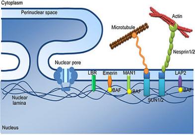
Structural support is provided to the nuclear envelope along the inner surface of the nucleus by a special mesh-like lining called the nuclear lamina, which binds to chromatin, integral membrane proteins, and other nuclear components. The nuclear lamina is also thought to play a role in directing materials inside the nucleus toward the nuclear pores for export and in the disintegration of the nuclear envelope during cell division and its subsequent reformation at the end of the process. Another intermediate filament network is located outside the nuclear membrane and is not organized in such a systemic way.

Fig. 6

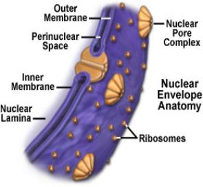
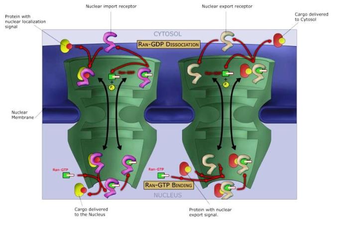
There is high volume traffic through nuclear pores. RNA and ribosomal subunits must be constantly transferred from the nucleus where they are made to the cytoplasm and histones, gene regulatory proteins, DNA and RNA polymerases, and other substances required for the nucleus must be imported. An active cell can synthesize about 20,000 ribosome subunits per minute, and at certain points in the cell cycle, as many as 30,000 histones per minute are required. For such a huge number of molecules to pass through the nuclear pores (below), they must be highly efficient at selectively allowing passage.

Fig. 7

Chromosomes

The genetic code is written by the pairing of four nucleotide bases which are represented by the color rungs of the DNA helical ladder in Figure 8. Adenine (A) forms a base pair with thymine (T) and guanine (G) forms a base pair with cytosine (C). In RNA, thymine is replaced by uracil (U). The order of each of the three rungs form a codon for one of 20 amino acids, like three letter words for each amino acid, self-similar to the three Cycles of System 4. The sequence allows an unlimited variety of proteins.

Cell division and gene expression is controlled in part by epigenetic processes, the series of actions that affect the protein spools around which the DNA is tightly bound, rather than encoded in the DNA sequence itself. Those spools are built of histone proteins, and chemical changes to them can either loosen or tighten their interaction with DNA. Epigenetics alter the readout of the genetic information, in some cases ramping a gene's expression up or down.

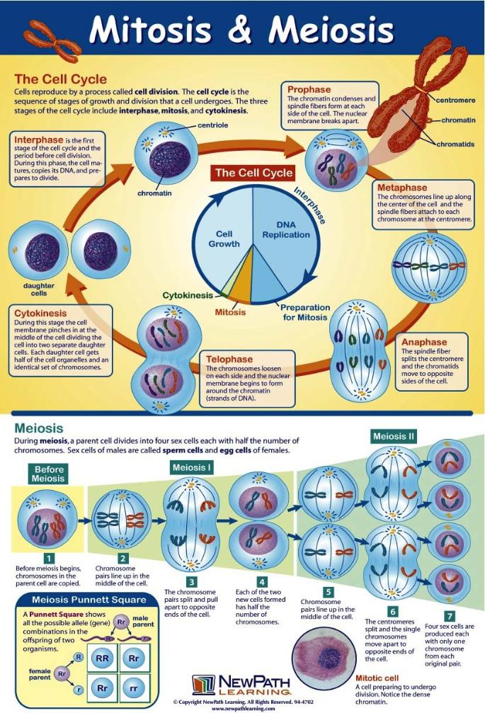
Prior to cell division, DNA replication of chromatin takes place. Both DNA strands serve as templates for the reproduction of the opposite strand. Replication begins at specific places in the genome, called origins. As the DNA unwinds at the origin, the synthesis of new strands forms at a replication fork. In addition to DNA polymerase, other enzymes at the fork help to start and continue the DNA synthesis. The two identical copies of chromatin are joined at their centromeres, then the chromatin strings become densely folded into tightly wound chromosomes as in Figure 8. The Microtubule Organizing Center (MTOC) has two centrioles that replicate. One such complex migrates to opposite poles of the cell to generate the mitotic spindle as the nuclear envelope disappears. Spindle microtubules from each pole attach to the centromeres and move the two identical strings of chromatin apart as the cell divides.


In interphase, each DNA chromatin string is strung out to facilitate transcription of RNAs. Prior to cell division, it replicates into two identical chromatin strings joined at their centromeres. They become densely packed into distinct chromosomes for cell division. The diagram illustrates the folding of the top string of chromatin. The bottom string folds in an identical way and the two are joined at their centromeres. During mitosis, microtubules attach to the centromere from opposite poles and move the two identical chromatids apart. The two daughter cells thus receive identical copies of chromatin.

Fig. 8 [Above]

Mitosis and Meiosis

Fig. 9

The Nucleolus

The nucleolus makes the ribosomes that assemble amino acids into protein. The nucleolus is formed around specific genetic loci called nucleolar organizing regions (NORs) composed of tandem repeats of rRNA genes in several different chromosomes. In cell division, NORs disappear then reappear after.

Transcription of the ribosomal gene yields a long precursor molecule which requires further processing. RNA-modifying enzymes are brought to their recognition sites by guide RNAs, which bind these specific sequences. A variety of other small RNAs are involved in processing. Once the rRNA subunits are processed, they are ready to be assembled into larger ribosomal subunits. However, an additional rRNA molecule is necessary, which is transcribed in the nucleoplasm, then relocates to the nucleolus for ribosome assembly. This assembly requires ribosomal proteins as well.

The required mRNA to make them is transcribed and processed in the nucleoplasm, then exported for protein synthesis on free ribosomes in the cytoplasm. The mature r-proteins are then "imported" back into the nucleus, and finally, the nucleolus. Association and processing of rRNA and r-proteins result in the formation of the small and large subunits of the complete ribosome. These are exported through the nuclear pore complexes to the cytoplasm, where they remain free or become associated with the endoplasmic reticulum.

A continuous chain between the nucleoplasm and the inner nucleolus exists through a network of nucleolar channels. In this way, macromolecules are easily distributed throughout the nucleolus.

Ribosomes and RNA

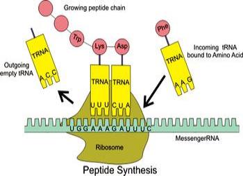
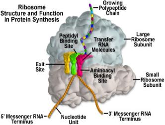
Ribosomes are machines in the Production Department of the cell. They assemble amino acids into protein using messenger Ribonucleic Acid, mRNA, and transfer tRNA. Protein is assembled from 20 kinds of amino acids in specific sequences. The DNA codon sequence in genes is copied and processed into an mRNA for each kind of protein. Ribosomes then read the three letter codon information for each amino acid in the mRNA sequence, while tRNA brings each amino acid specified by each successive codon to the ribosome for attachment to the growing amino acid (polypeptide) string that will constitute the finished protein. Transfer RNA is specific to each DNA codon and to its related amino acid. The ribosome moves along the mRNA strand, reading its codon sequence and producing a chain of amino acids. Ribosomes can attach in series to an mRNA strand. These polysomes make protein in parallel.

Ribosomes are divided into two subunits. The smaller subunit binds to the mRNA, while the larger subunit successively attaches to each tRNA and adds its amino acid to the polypeptide string. When a ribosome finishes reading a compete strand of mRNA, the two subunits split apart until they reattach to another mRNA. Ribosomes have also been classified as ribozymes because the ribosomal RNA seems to be most important for the enzyme peptidyl transferase activity that links amino acids together.

Fig. 10

Initiation of Transcription

Eukaryotic RNA polymerase (RNAP) transcribes the RNA from its gene. A collection of proteins called transcription factors (in color) mediate the binding of RNA polymerase and the initiation of transcription. Only after certain transcription factors are attached to the DNA promoter sequence for the gene, does the RNA polymerase bind to it.

Transcription Elongation of Polypeptide (Protein) Strand

The two strands of DNA run in opposite directions as designated by 3' and 5' ends (numbered nucleotide carbon atoms). One strand of the DNA, the template strand (or noncoding strand), is used as a template for RNA synthesis. As transcription proceeds, RNA polymerase traverses the template strand and assembles base pairing with the DNA template to create an RNA copy. This produces an exact copy of the coding strand (except that thymines are replaced with uracils, and the nucleotides are composed of a ribose (5-carbon) sugar where DNA has deoxyribose (one less oxygen atom) in its sugar-phosphate backbone).

Termination of Transcription

Transcription termination in eukaryotes involves cleavage of the new RNA transcript from the template followed by template-independent addition of Adenines at its new 3' end, in a process called polyadenylation. The completed mRNA strands are exported to the cytoplasm where they act as templates for protein synthesis at ribosomes. The exported tRNAs are processed and folded in a specific way to attach specific amino acids for assembly.

Fig. 11 [Above]

In large corporations, the Engineering Department prepares the plans for making specific products for sale and for maintaining the company infrastructure in a proper working condition. In all host cells, the same DNA plans have been prepared by evolution, so this subsumed engineering function identifies how, when, and where to read them. However, the amount of each product to be made for export depends on current market demand as assessed by the Sales Department. Market needs as interpreted via cell signaling is associated with the cell's overall organization with respect to the environment. Sales balances input signals with an output response that involves the kind and quantity of RNAs produced as well as the number of ribosomes needed. Starvation, insufficient nutrients and other factors selectively inhibit cell signaling pathways so signaling concerns the overall cell Organization Department.

The kind of RNA's needed for specific proteins and enzymes depends upon the kind of cell and signaling pathways that initiate the needed RNA transcription. The frequency of signals will affect how much RNA transcription is needed. This depends on the concentration of specific ligand messengers, the number of parallel pathways, and the pre-initiation complex in the nucleus. The pre-initiation complex contains: 1. Core Promoter Sequence, 2. Transcription Factors, 3. DNA Helicase, 4. RNA Polymerase, 5. Activators and Repressors. Hundreds of protein transcription factors have been identified that likewise need to be synthesized or activated. The helicase enzymes unzip the relatively weak hydrogen bonds that join the two DNA strands to make transcription possible, then the RNA polymerase assembles the RNA strands. This identification of the appropriate gene and the related initiation factors are an Engineering function.

Unlike DNA replication, mRNA transcription can involve multiple RNA polymerases on a single DNA template and multiple rounds of transcription, so many mRNA molecules can be rapidly produced from a single copy of a gene. The nucleolus can be churning out ribosomal sub-units at the same time. This corresponds to a Sales function with respect to products for export, but remains an Engineering function as it relates to cell maintenance. Protein synthesis is a Production Department function. It begins when the mRNA attaches to ribosomal subunits while tRNA brings the amino acids in the sequence coded for.

The Production process is similar both for protein export and cell maintenance except that, in the former case, the signaling cascade is commonly initiated from signal receptors embedded in the cell membrane, whereas in the latter case, calcium ion channels play a central role in regulating signaling. Synthesis for export occurs at ribosomes that attach to the endoplasmic reticulum (ER) and thread the polypeptide chain inside the ER lumen. Synthesis for cell maintenance occurs either at free ribosomes in the cytosol or ER ribosomes that release protein to the cytosol. The smooth ER lacks ribosomes. It metabolizes carbohydrates, makes membrane lipids, steroids, and processes signal receptors for inclusion in vesicles and the plasma membrane.

The Endoplasmic Reticulum

The endoplasmic reticulum (ER) is an assembly line in the Production Department where many of the ribosomal machines do their work, with many others working freely in the cystosol. There may be a million or more ribosomes in a cell transcribing a couple thousand mRNA types depending on cell type and current needs. A notable exception is the 25 trillion or so red blood cells in the body that lack a nucleus, mitochondria, and ER and have no ribosomes. The bone marrow takes about 7 days to make them from stem cells, yet turns out over 5 million an hour.

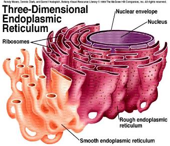
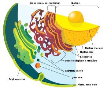
The ER consists of an extensively folded continuous labyrinth of flattened sacs and tubes that extend throughout much of the cytoplasm. It consists of areas of rough ER together with areas of smooth ER. The rough ER is densely peppered with ribosomes that give it its rough appearance. The interior compartment inside the ER membrane is called the lumen and is separate from the cytoplasm, although it is continuous with the lumen inside the double nuclear membrane.

Fig. 12

When ribosomes first begin to synthesize protein, they are not attached to the ER. If a signal sequence of amino acids is attached to the leading edge of the polypeptide string coded for by the mRNA, there is a signal recognition particle (SRP) in the cytosol outside the ER that guides the signaled end of the string to enter a transporter protein channel embedded in the ER membrane. The SRP docks on a signal receptor beside the transporter channel. Once the string being assembled is entering the ER lumen, the SRP detaches from the protein channel to shuttle back for another, and an enzyme in the ER lumen removes the signaled sequence on the end of the polypeptide string. When the translation of the RNA sequence into protein has completely threaded inside the ER, the small and large sub units of the ribosome disassociate until the leading end of another RNA sequence attaches to reunite them.

The sections of smooth ER are more tubular and lack ribosomes. Within its lumen, some proteins synthesized in the rough ER are modified. Phospholipids, steroids and fatty acids are synthesized there, some carbohydrates are metabolized and toxic substances are rendered inert. Signaling receptors are attached to proteins destined to be integral in new sections of membrane that bud off and are transported to peroxisomes, the Golgi apparatus, and the plasma membrane. ER proteins receive chemical address tags for specific purposes in the lumen of the ER through a series of chemical reactions. The mitochondrial outer membrane can associate with the ER membrane, in a structure called MAM (mitochondria-associated ER-membrane). This is important in ER-mitochondria calcium signaling and involved in the transfer of lipids between the ER and mitochondria.

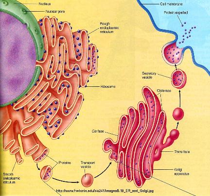
The ER processes proteins that are further processed in the Golgi apparatus generally for export from the cell or for use in vesicles such as lyosomes. Ribosomes that do not produce proteins for export via the ER to Golgi pathway synthesize proteins that remain soluble in the cytosol for use within the cell.

Fig. 13

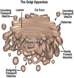
The Golgi apparatus (GA) stores, modifies, and packages proteins primarily for export. It consists of a stack of saucer shaped sacks each enclosed in a lipid membrane. Vesicles containing proteins bud off from the ER and move through the cytoplasm to fuse with what is called the cis region of the GA nearest the ER. They disgorge their contents into the lumen of a GA sack. Other small vesicles move from sack to sack in the stack towards the "trans" region of the GA where vesicles package the end product. In the process, the vesicle is addressed to a specific destination, either for export, storage vesicles, or for use in lyosomes. The GA exports some proteins constantly and retains others for use at appropriate times. As membrane is used for vesicle formation, more can be synthesized by the ER. The ER, GA, and nuclear membranes are disassembled during cell division and reassembled after.

As part of the Production Department, the GA is a product processing and warehousing center for the export of proteins, lipids, and other large molecules. Much of our body is made up of exported material, including antibodies, hormones, digestive enzymes, growth factors, and much of the material that makes up bone, cartilage, skin, and hair. A major processing activity is the adding of sugar molecules to form glycoproteins. In cells such as mucous secreting cells, the amount of carbohydrate added far exceeds the amount of protein. Hormones and neurotransmitters are too small to be synthesized directly by ribosomes, so large precursor proteins are cut up as they pass through the GA. The lipid membranes of export vesicles contain integral signaling receptor proteins that become part of the cell membrane.

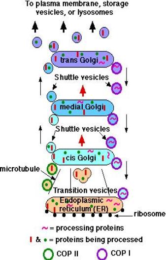
• Transition vesicles pinch off from the surface of the ER carrying integral membrane proteins, soluble proteins awaiting processing, and enzymes

• Pinching off requires that the vesicle be coated with COPII (Coat Protein II)

• The transition vesicles move toward the cis Golgi on microtubules

• As they do so, their COPII coat is removed and they may fuse together forming larger vesicles

• These fuse with the cis Golgi

• Sugars are added to proteins in small packets so many glycoproteins have to undergo a large number of sequential steps of glycosylation, each requiring its own enzymes

• These steps take place as shuttle vesicles carry the proteins from cis to medial to the trans Golgi compartments

• At the outer face of the trans Golgi, vesicles pinch off and carry their completed products to various destinations

• The movement of cisternal contents through the stack means that essential processing enzymes are also moving away from their proper site of action

• Using a variety of signals, the Golgi separates the products from the processing enzymes that made them and returns the enzymes back to the endoplasmic reticulum

• This transport is also done by pinching off vesicles, but the inbound vesicles are coated with COPI (Coat Protein I)
Fig. 14

The Cytoskeleton

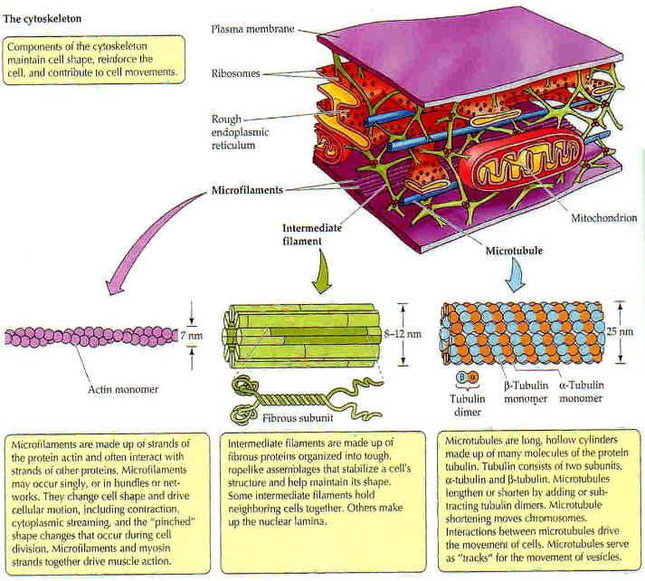
The cytoskeleton consists of a dynamic set of microfilaments, intermediate filaments, and microtubules that position organelles and determine the shape of the cell essential to organ tissues. They also provide for various types of cell movement and some act as tracks for motor proteins that help a cell move or that move molecular complexes and vesicles within a cell. Motor proteins such as myosin have legs about 25 nanometers long that attach and detach from fiber proteins and typically take 7.2 nanometer strides along actin fibers. They can walk about 1 micrometer a second, so they can move cargo long distances inside a cell in seconds and completely across a cell in about a minute. A typical cell may be 50-100 micrometers in diameter or 2,000 to 8,000 times the diameter of a ribosome. Since volume varies as the cube of the radius, a ribosome is less than a billionth the volume of a typical cell.

The cytoskeleton is concerned with the structural Organization of events in each Cell as it relates to Organs and Host and as this subsumes Organelles and Enzymes within each Cell. Microfilaments made of two G-actin protein chains form a double helix. They are involved in muscle contraction and localized changes in the shape of other cells such as the pinching contractions in cell division. There are at least five distinct but similar types of intermediate filaments that play more static roles lending a degree of rigidity to the cell and holding organelles in place. Microtubules radiate from the microtubule organizing center (MTOC). They are long hollow un-branched cylinders that can change length rapidly. They are involved in changes in cell shape and also serve as tracks to guide motor molecules that transport vesicles, which in turn can induce cytoplasmic streaming (currents in the cytosol). Microtubules are essential in cell division and they are primary structures in centrioles, flagella, and primary cilia. The network of cytoskeleton filaments restricts the diffusion of large particles in the cell. Particles larger than the size of a ribosome are excluded from parts of the cytosol around the cell edges and next to the nucleus. These "excluding compartments" may contain a much denser actin fiber meshwork than the remainder of the cytosol, influencing the distribution of larger complexes by excluding them from some areas and concentrating them in others.

Fig. 15

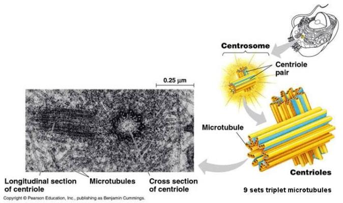
Centrioles and the MTOC

The microtubule-organising center (MTOC) is a structure found in eukaryotic cells from which microtubules emerge. MTOCs determine the organization of eukaryotic flagella and cilia and the organization of the mitotic and meiotic spindle apparatus that separates chromosomes during cell division. In animals, the centrioles are important aspects of MTOCs. The mother centriole acts as the basal body from which the primary cilium grows into a projection from the main body of the cell like a tiny antenna that is in communication with other cells.

During interphase, animal cells have one centrosome, usually located near the nucleus. The centrosome has a pair of centrioles, except for cells that do not have a nucleus (red blood cells). Microtubules are anchored with their "minus" ends in the centrosome, and because microtubules dissociate preferentially at this end, this anchoring has a stabilizing effect, allowing microtubules to grow very quickly. The polarity of the microtubules is important for membrane bound transport vesicles, since the motor proteins kinesin and dynein typically move preferentially in either the "plus" or "minus" direction, respectively, along a microtubule, allowing vesicles to be directed to or from the endoplasmic reticulum and Golgi apparatus. The centriole pair is always oriented at right angles to one another.

Fig. 16

Primary Cilium

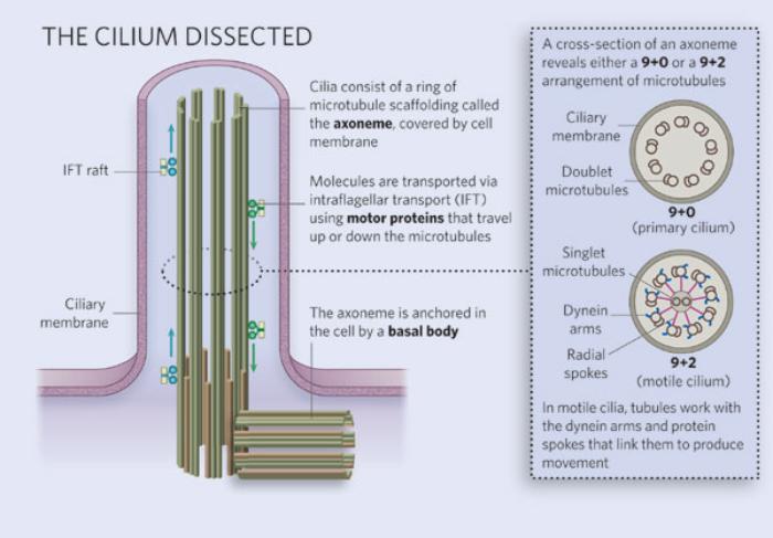
Most cells in the body have one non-motile primary cilium distinct from cilia in epithelia cells that beat synchronously. Carefuly examination of kidney tubule cells showed that the primary cilia bend when exposed to moving liquid as is the case in working kidneys. This discovery showed that primary celia were acting as sensors called mechanoreceptors, responding to flow by opening calcium channels and allowing a rapid influx of calcium ions into the cells. Primary cilia also respond to chemical signaling, light, osmolarity, temperature, and gravity.

Primary cilia are cetnral to a wide variety of receptor functions and signaling. In addition to simple signal transduction, they have an important role in a variety of processes, including development, differentiation, and some types of memory. They act as antennae that synchronously Organize diverse Cell functions in Organs of the Host human being. This overall integration of Cell, Organs, and Host subsumes the Organization of each Host Cell as it relates internally to its Organelles and Enzymes.

The basal body of each primary cilium is the mother centriole with a 9×3 structure. The Primary Cilium grows on top of it with a 9×2 structure with a transition zone between. The 9×2 structure is consistent with the 20 Term transformations of System 5, except that the two central microtubules that are present in motile cilia are absent in primary cilia. Motile cilia in organ tissues, such as the brain ventricles and lungs, beat synchronously indicating their role as channeling synchronous energies between cells of the same organ tissue type. In non-motile primary cilia, their absence indicates that the synchronous function of Cells in Organs of the Host is not delegated at the level of the cell. The primary cilia act as antennae to synchronously integrate all body functions in accord with the needs of the human Host.

Fig. 17

The Cytosol, Metabolons, Protein Vaults, and Proteosomes

Apart from the nucleus of the cell, the other cell contents constitute the cytoplasm of the cell. The cytosol is the intracellular fluid in the cytoplasm. It is involved in a variety of functions that include signal transduction, cytokinesis, and transport of metabolites from the production site to the place where they are used. Major metabolic pathways such as the pentose phosphate pathway, glycolysis, protein synthesis, and gluconeogenesis occur within the cytosol. The cytosol is mostly water with many large water-soluble molecules, proteins, and a host of small molecules and ions. It includes protein complexes, concentration gradients, calcium waves, cytoskeletal sieving, and protein assemblies and compartments.

The cytosol has thousands of enzymes involved in metabolism. Many of the newly synthesized proteins remain in the cytosol if they lack a signal for transport. About 70% of the cytosol is water with dissolved nucleic acids, enzymes, amino acids, lipids, carbohydrates, ions, and nutrients that are moved within the cell with the help of cytoplasmic streaming. Proteins are moved to their appropriate location based on transport signals attached to them. The human genome can code for about 200,000 proteins.

Metabolons

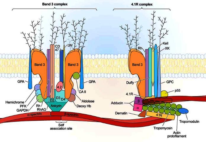
The cell has multi-step enzymatic reactions that are often facilitated by large protein assemblies called metabolons. A metabolon is a temporary structural-functional complex formed between sequential enzymes of a metabolic pathway. It is held together by non-covalent interactions. They may also be held together by structural elements of the cell such as integral membrane proteins and proteins of the cytoskeleton. Metabolons facilitate passing the intermediary metabolic product from one enzyme directly onto the active site of the consecutive enzyme of the metabolic pathway. The Kreb's Cycle (Citric Acid Cycle) is an example of a metabolon which facilitates substrate channeling. During the functioning of metabolons, the amount of water needed to hydrate the enzymes is reduced and the enzyme activity is increased. Protein assemblies associated with red blood cell integral membrane proteins are illustrated in Figure 18.

Fig. 18

Vaults

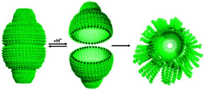
Ribonucleoproteins self assemble into nano-sized hollow structures called vaults. The internal chamber is large enough to contain a ribosomoe and may be involved in their transport. There are about 10,000 vaults in a typical human cell. Despite not being fully elucidated, vaults have been associated with nuclear pore complexes. It is thought that they transport molecules, such as mRNA, from the nucleus to parts of the cytoplasm and that they play a role in protein synthesis in the cytosol. Vaults are actively transported along microtubules within nerve axons to the nerve terminal suggesting that microtubule transport would enable vaults to shuttle cargo directionally to specific locations in the cell. Vaults are also indicated in the nuclear targeting of steroid hormone receptors, most notably the estrogen receptor, and hence may play a role in the signal transduction of steroid hormones.

Vaults dissociate into halves at low pH. At low pH, the 234 acidic residues at the vault interface become neutral, leaving a highly electropositive charge and inducing the disassembly of the vault particle by charge repulsion (Figure 19). At higher pH, the aspartate and glutamate residues recover their acidic state and re-establish the electrostatic interactions, allowing the re-association between the two vault halves. Subsequently, the hydrophobic interactions contribute to stabilize the locked conformation of the particle. This allows for the encapsulation and release of contents which can be locally affected by calcium signaling and other factors.

Fig. 19

Proteasomes

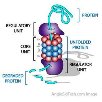
Proteasomes are large protein complexes in the nucleus and cytoplasm. They degrade unneeded, damaged or misfolded proteins by employing protease enzymes to break peptide bonds. The degradation process yields peptides of about seven to eight amino acids long, which can then be further degraded into amino acids and used in synthesizing new proteins. Figure 20 is highly simplified.

A proteasome is a cylindrical complex containing a "core" of four stacked rings around a central pore that contains six protease active sites. Target protein must enter the central pore before it is degraded. Proteins are tagged for degradation with a small protein called ubiquitin, a reaction that is catalyzed by enzymes called ubiquitin ligases. Once a protein is tagged, this signals other ligases to attach additional ubiquitin molecules. The result is a polyubiquitin chain that is bound by the proteasome and feeds through a gate into the central pore where the chain is degraded. Since the protease active sites are inside the barrel of the proteosome, other proteins in the cytosol are spared enzyme degradation.

Fig. 20

Peroxisomes

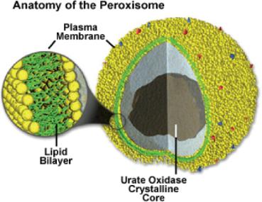
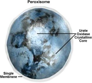
Peroxisomes contain a variety of enzymes to rid the cell of toxic substances, in particular, hydrogen peroxide (a byproduct of cellular metabolism). They convert the hydrogen peroxide to water, rendering the potentially toxic substance safe for release back into the cell.

Some types of peroxisomes, such as those in liver cells, detoxify alcohol and other harmful compounds by oxidizing bonded hydrogen to make water. Others are more important for their ability to initiate the production of phospholipids used in the formation of membranes. In order to carry out their activities, peroisomes use oxygen. A major function of the peroxisome is the breakdown of very long chain fatty acids through beta oxidation to make medium chain fatty acids, which are shuttled to mitochondria where they are eventually broken down to carbon dioxide and water. Peroxisomes initiate the formation of plasmalogen which is essential to the myelin sheath in nerve fibers. Peroxisomes also play a role in the production of bile acids for the absorption of fats and fat-soluble vitamins, such as vitamin K.

Peroxisomes can be derived from the ER and they replicate by fission. Peroxisome matrix proteins are made by ribosomes in the cytoplasm prior to import. Specific amino acid signal sequences (PTS1 and PTS2 attached during translation) target the proteins to be imported into the organelle. There are at least 32 known peroxisomal proteins, called peroxins, which participate in the process of peroxisome assembly. The protein receptors, the peroxins PEX5 and PEX7, accompany their cargoes (containing a PTS1 or PTS2 amino acid sequence respectively) all the way into the peroxisome where they release the cargo and then return to the cytosol. ATP hydrolysis is required to energize this shuttling operation. Peroxisomes can synthesize some of their own membrane lipids, but also obtain some from the ER. Peroxisomes can increase in number by dividing once they grow large enough.

Fig. 21

Lyosomes

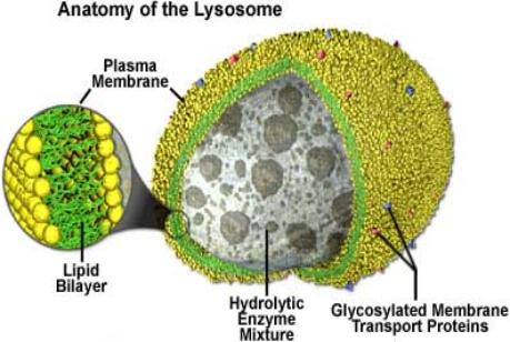
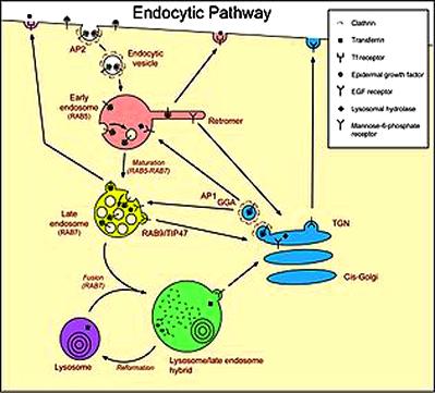
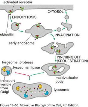
Lyosomes combine two vesicles. One buds off from the Golgi apparatus and contains about 40 or more digestive enzymes. The other engorges food and other particles from outside the cell by endocytosis (similar to Figure 23) which buds off a phagosome vesicle from a pocket in the cell membrane to the inside of the cell. The two vesicles then combine into a secondary lyosome and the enzymes rapidly digest the contents of the phagosome. The products of digestion diffuse into the cytoplasm through the single membrane of the lyosome. The lyosome then fuses with the cell membrane to disgorge the undigested or waste particles outside the cell. The digested products provide fuel molecules and raw materials for other cell processes. By containing the digestive enzymes within the lyosomes, they are prevented from damaging the rest of the cell. Phagosomes can be compared to Production Department purchasing of raw materials that require processing before they can be employed in product manufacture. Enzymes are the workers. They catalyze reactions without being chemically altered.

The lyosome enzymes are synthesized on RER and further processed in the Golgi apparatus. They are also involved in autophagy. During this process, old worn-out parts of the cell, like old mitochondria, are digested. So, lysosomes are also involved in the recycling of some old cell contents.

Fig. 22

Vesicles

Vesicles are small bi-lipid membrane enclosed sacks that can store or transport substances. Most vesicles have specialized functions depending on what materials they contain. Vesicles can fuse with the membranes of other organelles and with the cell membrane. This allows them to store, transport, digest particles and discharge waste. Vesicles are involved in metabolism, transport, buoyancy control, enzyme storage, and they also act as chemical reaction chambers.

Transport vesicles can move molecules between locations inside the cell, such as proteins from the rough ER to the Golgi apparatus. Secretory vesicles contain materials that are to be excreted from the cell, such as waste, neurotransmitters, hormones, and materials for the extracellular matrix.

A multivesicular body (MVB) is a membrane-bound vesicle containing a number of smaller vesicles as illustrated by the endosome as it matures in Figure 23. Small vesicles bud off internally in the endosome to compartmentalize and sort its contents for redistribution.

The vesicle coat sculpts the curvature of a donor membrane. It selects specific cargo proteins by binding to complex sorting signals. In this way, the vesicle coat collects selected membrane cargo proteins into nascent vesicle buds. Surface markers called SNAREs identify the vesicle's cargo, and complementary SNAREs on the target membrane act to cause fusion of the vesicle and target membrane.

Fig. 23

Mitochondria

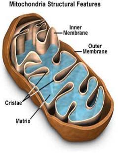
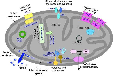
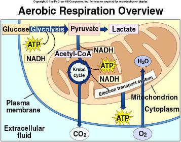
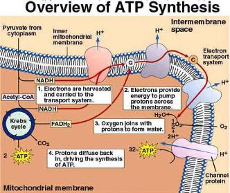
Mitochondria are small, semi-autonomous organelles that possess their own DNA although they require the cooperation of the nuclear DNA also. They are like a Treasury Department that funds most of the energy for the operation of the other cell machinery and related chemistry. The utilization of food, such as glucose, begins in the cytosol. The pyruvate fuel molecules resulting from glycolysis of glucose enter mitochondria whose primary function is to convert their energy (using oxygen in a process called cellular respiration) into molecules called ATP (adenosine triphosphate), a form of energy currency that the cell can use. Like money in a market economy, ATP (adenosine tri-phosphate) participates in chemical processes that require energy by exchanging phosphate ions, a process called phosphorylation. Enzymes called kinases phosphorylate, and phosphatases dephosphorylate. Mitochondria have a double membrane. The smooth outer membrane offers little resistance to substances diffusing through it both ways, unlike the inner membrane which is highly selective.

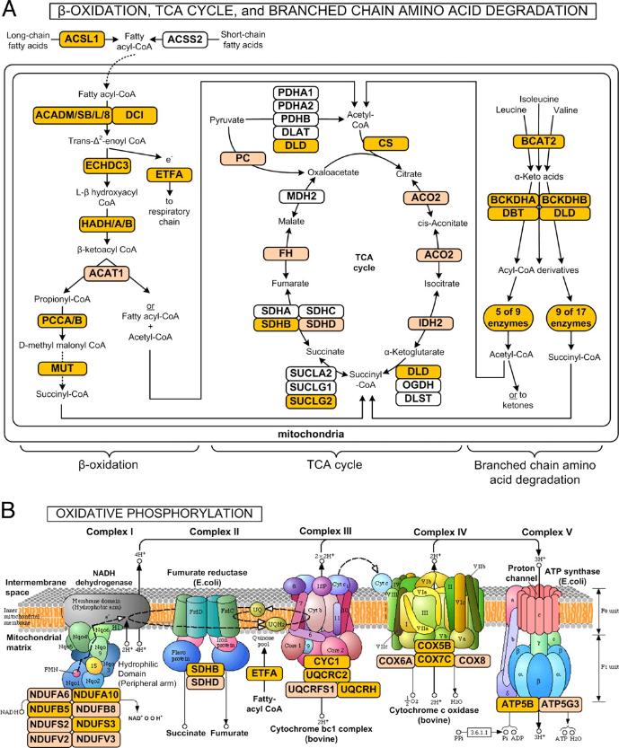
The inner membrane folds inward in many places to form shelf-like "cristae" that greatly increase its surface area. This membrane contains many large protein complexes in the electron transport chain that results in the cellular respiration of ATP (A more detailed diagram is in the Appendix). It also exerts control over what enters or leaves the region inside called the matrix. The matrix contains ribosomes, enzymes, and two to ten copies of its circular DNA that allow it to make some of the proteins it needs. Most of the many hundreds of proteins it needs are synthesized in the cytosol and imported. The mitochondrial genome codes for 13 sub-units of respiratory complexes used in the Krebs (citric acid) cycle. Another 22 genes code for tRNA and two code for rRNA. Some nuclear RNAs maek their way into mitochondria. Cells may have up to many thousands of mitochondria, depending on their energy needs. Mature red blood cells have none, while muscle and nerve cells have many.

The main roles of mitochondria are to produce ATP through respiration, and to regulate cellular metabolism. The central set of reactions involved are collectively known as the TCA cycle, citric acid cycle, or the Krebs Cycle. The glycolysis of glucose in the cytosol produces pyruvate which is imported into mitochondria and oxydized as feedstock for the TCA cycle. Mitochondria can store and release calcium ions and regulate an array of reactions in the cell including signal transduction. The endoplasmic reticulum (ER) is the most significant storage site of calcium, and there is a significant interplay between the mitochondrion and ER with regard to calcium. Mitochondria thus play a critical role in cell signaling events, in cell responses to a multiplicity of physiological and genetic stresses, inter-organelle communication, cell proliferation, and cell death. This may be summarized as follows:

• Storage of calcium ions. Regulation of the membrane potential.
• Heat production
• Apoptosis (programmed cell death)
• Calcium signaling (including calcium-evoked apoptosis)
• Cellular proliferation regulation
• Regulation of cellular metabolism
• Certain heme synthesis reactions
• Steroid synthesis

Some functions are performed only in specific types of cells. For example, mitochondria in liver cells contain enzymes that allow them to detoxify ammonia, a waste product of protein metabolism.

Fig. 24

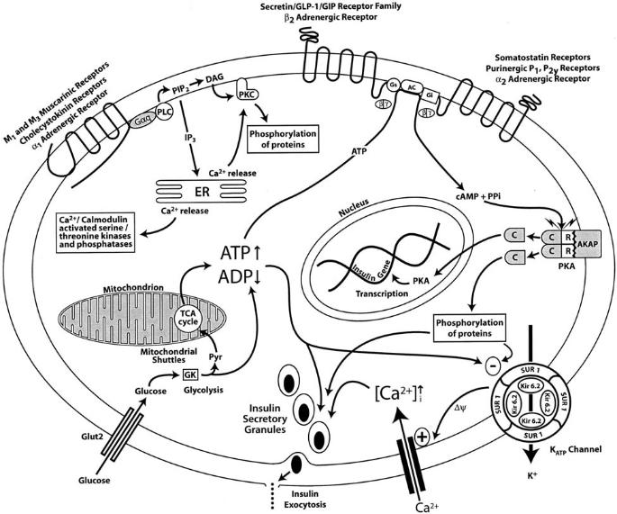
Insulin Regulation in Pancreatic Cells

In healthy individuals, insulin is secreted by pancreatic cells into the bloodstream after a meal in response to elevated blood sugar.

Figure 25 is simplified to show some of the main pathways during glucose-induced insulin secretion in the β-cells of the pancreas. Glucose enters via the glucose transporters. It is phosphorylated and this determines the rate of glycolysis and the rate of pyruvate generation for entry into the mitochondira. Therefore, when blood glucose is high, the rate of glycolysis will increase. In the mitochondira, pyruvate is teh substrate for oxidation in the TCA (citric acid) cycle that ultimately activates the electron transport chain. (Not shown. See Figure 24 and the Appendix.) The electron transport chain generates ATP which is exported from mitochondria to the cytoplasm. The incrase in the cytoplasmic ATP/ADP ratio closes the KATP channels and depolarizes the plasma membrane, allowing the opening of voltage-dependent calcium channels and a rapid influx of Ca2+. The increase in cytosolic Ca2+ is the main trigger in glucose-dependent insulin secretion from primed secretion vesicles.

Intracellular calcium (Ca2+) can also be released from sequestered stores in organelles. Activation of Gα (by α1 or muscarinic receptor ligands, for example) leads to hydrolysis of phosphatidyl inositol biphosphate to produce inositol triphosphate (IP3) which opens calcium channels in the ER releasing Ca2+. DAG activates protein kinase C, at least partly by sensitizing it to CA2+. Activation of the Ca2+/calmodulin kinases also occurs with the rise of Ca2+. The Gs protein (a guanine nucleotide-binding protein) is coupled to the subfamily B G-protein-copuled receptors and β2 adrenergic receptors and leads to activation of adenylyl cyclase and subsequently of PKA. Activated Ca2+/calmodulin kinases, PKC, and PKA can lead to phosphrylation of a myriad of proteins throughout the β-cell associated with the insulin secretory vesicles, the ion channels, and the cytoskeletal structure. Phosphorylation and dephosphorylation reactions initiated through these G-coupled pathways also ultimately regulate transcription of genes involved in the regulation of insulin secretion. Insulin secretion is part of a subsuming Treasury function as it relates to Organs and Host. It budgets energy use in the body.

Fig. 25

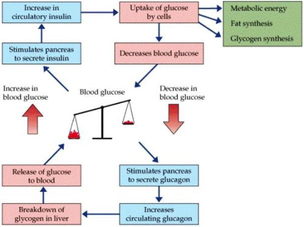
Insulin regulation is linked to energy use and storage throughout the body as simplified in Figure 26. High blood sugar levels in excess of immediate energy needs can be converted for storage as long polymer chains of glucose called glycogen. This takes place mainly in the liver, but also in muslces, and to a small degree in some other cells. Fatty acids are also stored in the liver, muscles, and fat cells. When blood sugar is low, glucagon signals adipocytes (fat cells) to convert triglycerides into free fatty acids. Serum albumin in the blood binds free fatty acids and transports them to organs such as muscle and liver for oxidation to ATP when blood sugar is low. This storage and recall of energy is a kind of Treasury accounting for energy savings and expenditures.

Fig. 26

Typical Glucose Metabolism in Muscle Cells

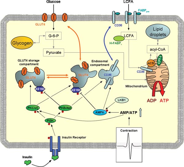
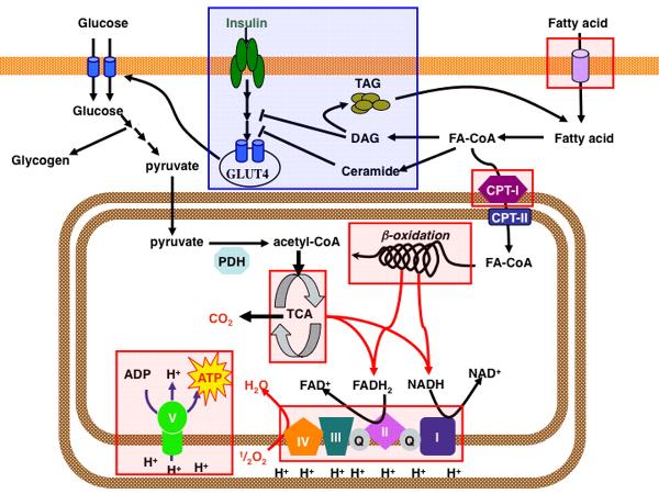
Signaling pathways and the regulation of cardiac metabolism are shown simplified in Figure 27. Glucose and long-chain fatty acids (LCFA) are the major substrates for cardiac energy production. Their import is increased after translocation of specific transport proteins (mostly GLUT4) to the sarcolemma (muscle cell membrane) in response to stimulation with insulin or during increased contraction. Glucose is either stored as glycogen or broken down to pyruvate in the cytoplasm. LCFA are taken up by trans-membrane CD36 (inserted as an integral transmembrane protein) and FABPpm (plasma membrane-associated Fatty Acid-Binding Protein). After import, LCFA is transported through the cytoplasm to the outer mitochondria membrane and converted by acyl-CoA synthetase (ACS) into acyl-CoA, which can be either esterified and stored in lipid (fatty acid) droplets or transported into mitochondira via CPT1. Finally, pyruvate and acyl-CoA are oxidized within mitochondria to generate adenosine triphosphate (ATP). Two independent stimuli lead to GLUT4 and CD36 translocation to the membrane from storage compartments, i.e. insulin stimulation and enhanced contractile activity.

Fig. 27

Effect of Insulin on Glucose Metabolism Summary

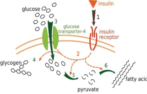
Insulin binds to its receptor (1), which starts many protein activation cascades (2). These include translocation of Glut-4 transporter to the cell membrane and influx of glucose (3), glycogen synthesis (4), glycolysis (5) and fatty acid synthesis (6). Glycolysis produces pyruvate which is the substrate for oxidative respiration in mitochondria.

Fig. 28

Notes on the Pancreas Gland

The pancreas is a dual-function gland, having features of both endocrine and exocrine glands. The part of the pancreas with endocrine function is made up of approximately a million cell clusters called islets of Langerhans. Four main cell types exist in the islets that can be classified by their secretion: α cells secrete glucagon (increase glucose in the blood), β cells secrete insulin (decrease glucose in the blood), δ cells secrete somatostatin (regulates/stops α and β cells), and PP cells secrete pancreatic polypeptide.

The pancreas as an exocrine gland helps out the digestive system. It secretes pancreatic juice that contains digestive enzymes that pass to the small intestine. These enzymes help to further break down the carbohydrates, proteins, and lipids (fats) in the digestive process. The pancreas receives regulatory innervation via hormones in the blood and through the autonomic nervous system. These two inputs regulate the secretory activity of the pancreas.

Cell Structure and Function as it Relates to the System

As noted above, the organization of a living cell parallels that of a large corporation. Cells demonstrate structural self-similarity to a corporation and to the six Particular Terms of System 4 and their transform sequences. When getting into the detailed specifics of how this works, it is first necessary to interpret the universal hierarchy in the context of the Cell as it relates to Organ and Host, and also in the subsumed context of how the Host cell relates internally to its Organelles and the Enzymes that catalyze chemical reactions. In the overview of the cell presented here we will not get into the specifics of how the four active interfaces define the meaning in each of the nine Terms, apart from identifying the hierarchy. The purpose of this article is to provide a general overview of how the System relates to the cell, using the analogy of a large corporation. Related articles address the structural dynamics of System 4 Terms as they relate directly to cell structures and their processes.

A large corporation may be viewed as the organized energy expenditure of its employees and equipment. It is an integrated energy pattern that is equated to monetary terms budgeted and accounted for in its financial reports. In a living cell, energy is the currency of exchange. There is an overriding archetypal energy pattern that directs all of its myriad cogs to mesh the way that they do. The pattern has taken a few billion years to evolve into human beings that can question and try to understand how the whole process works.

There is a self-similar pattern to all phenomenal experience that keeps recurring in every context. In other articles, it is shown how all corporations have six primary domains or departments that relate in polar pairs to provide polar insight into the Performance, Potential, and Commitment dimensions of its operations. It works the same way in the integration of the human nervous system as also demonstrated in other articles.

This conforms to the way that System 4 works, and it will be shown that it works in a self-similar way in a living Cell as it relates to Organs in a Host human being. This same intimate triad permeates phenomenal experience in different disguises. The Host cell also relates internally to its Organelles and their Enzyme catalyzed chemical reactions in a self-similar way. In other words, the chemical reactions throughout our bodies are orchestrated by subsumed triadic archetypal energy patterns that work behind the physical moleculear scenes. These are the animating energy patterns that make us alive.

Enzymes are complex proteins that catalyze specific reactions by their physical shape without their protein reacting chemically apart from exchanging phosphate groups that act like energy switches to turn them on or off by alternating their shape. Enzymes catalyze the chemical reactions by which Enzymes themselves are produced, so the whole process is a closed loop intimately linked with Organelles and the archetypal energy pattern of the Host Cell. Enzymes enable the many marriages of physical atoms and molecules that clothe our Host bodies so long as we inhabit the planet Earth. It should be noted that the energy patterns that regulate the intimate triadic relationships between Cells, Organs, and Host are not themselves of a chemically physical nature. They have evolved over hundreds of millions of years.

Terms of System 4

As indicated in the aboev review, cells demonstrate a structural self-similarity to the six domains of any corporation as prescribed by the six Particular Terms of System 4. There are only nine possible ways that four active interfaces (called Centers) can mutually relate with respect to a common inside and outside. Each of these nine ways defines the meaning implicit within each Term. How the Universal Terms integrate the transform sequences of the Particular Terms will be dealt with in separate articles.

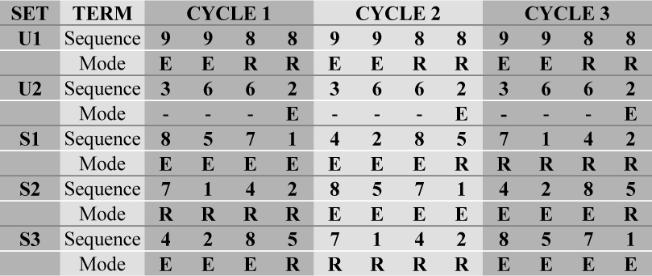
Apart from the Sales Term, each of the five remaining Particular Terms has both an Expressive and a Regenerative Mode. Since there are three Particular Sets of Terms transforming through the six Term sequence one Step apart, Terms 8, 7, and 4 synchronously alternate with Terms 1, 2, and 5. Some Terms are in the Expressive Mode while others are in the Regenerative Mode as in the following chart. This allows for spanning and integrating events in space and time through three Cycles of transformation. The vertical columns in the chart show the synchronous Terms in the three Particular Sets S1, S2, and S3. There are two Universal Sets each with their own transform sequence represented by U1 and U2.

Fig. 29

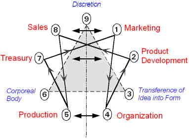
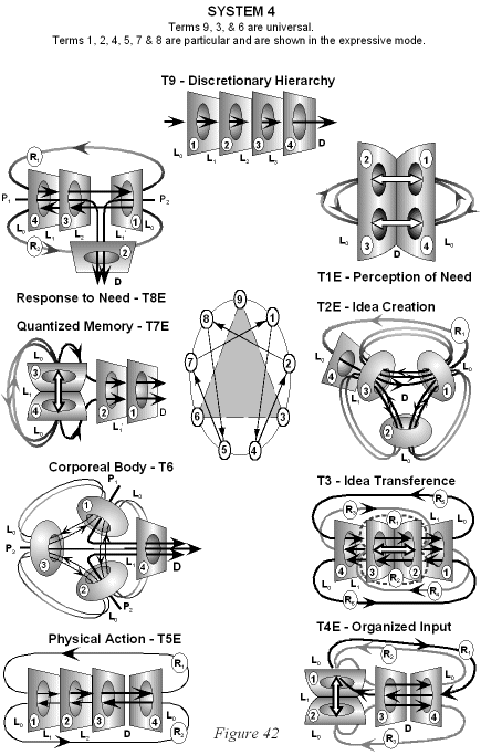
The Particular Term numbers 1, 4, 2, 8, 5, 7 correspond to the six primary domains of a corporation as illustrated in Figure 30. The analogous functions in a cell will be summarized below and subsidiary articles will expand accordingly. A detailed diagram of the nine Terms is in Appendix 3.

The Particular Terms of System 4 are shown in red as they apply to a business corporation. The flow of work transformation is shown by the connecting lines with arrows. The sequence transformations of the blue Universal Terms are not shown. The polar insights into the Performance, Potential, and Commitment dimensions of the company are shown by the three horizontal arrows respectively. The analogy is useful to understand cell organization and function.
Fig. 30

In Figure 30, it should be emphasized that Marking is distinct from Sales. Marketing concerns the forward vision of the organization and its preparedness to meet anticipated market needs and trends. Sales concerns meeting existing market demands with established products. Failure to recognize this distinction is the root cause of the recent financial meltdown. It should be noted that the Organization Department is usually called the Human Resources or Personnel Department. In a large corporation, Human Resources also concerns how the corporation is Organized to operate in response to anticipated Market needs. The corporation must be Organized and manned appropriately. This concerns the separate delegation of each of the six primary company departments in order to maintain polar insight into the creative dynamics of the corporation. Organization is not the arbitrary affair that it is often considered to be. It becomes quite complex in large organizations because the same six particular domains break out again within each of the primary departments.

The meaning in each System Term must be interpreted in context. In order to understand this, it is first necessary to interpret the universal hierarchy in the context of the Cell type as it relates to Organ tissues and Host human being as well to the Cell, Organelles, and Enzymes triad that directs molecular processes inside each cell. In the former case, the Particular Terms relate to collective events within each Cell as they collectively relate to Organs and the Host human being. In the latter case, the Particular Terms relate to chemical events catalyzed by specific classes of Enzyme teams associated with Organelles as these relate collectively to the overall integration of the host Cell. The universal Terms regulate their collective organization.

The Universal Hierarchy

Expressed in general terms, the universal hierarchy is IdeaKnowledgeRoutineForm

This is the Primary Universal Term T9. We exercise discretion accordingly. As a general example, if we have an Idea to build a snowman, this directs our Knowledge of how to do it, which directs our Routines of rolling snow into balls and stacking them, which results in the physical Form of the snowman. It works in a self-similar way in any creative enterprise, including natural biological processes. Discretion is a natural hierarchy.

Idea

The integrating idea of a living cell is a dynamic archetypal energy pattern that gives direction to all processes associated with the cell. The CEO of a business corporation performs this integrating function by appropriately balancing the three polar insights into the work performed in the six particular domains or primary departments of the corporation. One may think of this as an organized energy pattern expended by the structured work of employees in each of the six primary company domains or departments. It works in a similar fashion within a cell as it relates to organs and host. We may thus designate the Idea of the cell as Patterned Energy (PE). It includes the electronic environment of the cell membrane. It subsumes the myriad electro-chemical processes within the membrane and within the cell, including the intricately patterned exchange of phosphate ions. It is thus associated with, distinct from, the creative idea of synthesizing specific proteins within the cell, just as the integrating idea of a corporation is distinct from, but related to, the Product Development of specific products.

Knowledge

In a business organization, Knowledge is invested in the infrastructure of the corporation, in its manufacturing methods, buildings, equipment, and associated technical know-how. In a cell, this relates to the technology and equipment implicitly involved in the manufacture or synthesis of molecules needed both for its own regenerative needs as well as for export to the market environment of organs and host. We may thus designate this Organelle Knowledge (OK). Organelles in this context are not limited to membrane bound organelles. They include protein complexes such as ribosomes, vaults, metabolons, and proteosomes that perform specific functions. The nucleus is an organelle that contains the smaller organelles called the nucleolus and chromatin. Other organelles include the endoplasmic reticulum (ER), the Golgi apparatus (GA), mitochondria, lyosomes, vacuoles, vesicles, the MTOC with centrioles, primary cilia, the cell membranes, and the cyto-skeleton. This level concerns infrastructure cycles, including infrastructure renewal and cell replication.

Routine

In a business corporation, the Routine interface concerns product cycles that commit the resources of labor, equipment, and materials to creating products according to a schedule of needs specific to each of the comapny's six domains. It is a Supervisory Level of work that implicitly directs the Functional Level of making a physical product or service. In a cell, the supervisors are organized teams of enzymes that bring the raw materials and substrates together in specific sequences to perform chemical marriages and divorces. There are thousands of enzymes in a cell, but they work in supervised teams that are essential to each product cycle specific to each of the six domains that are evident within a cell. In the synthesis of enzymes and other proteins, they receive chemical address tags and peptide signals. Enzymes specific to various signaling and chemical pathways often associate into metabolons that organize the enzyme pathway more efficiently. Proteosomes contain enzymes that degrade protein. Enzymes are often packaged into vesicles and vaults that are transported by motor proteins that walk along microtubules to specific locations. For example, vesicles that transport neurotransmitters to the synapse contain a complement of enzymes. Also, sperm cells contain a vesicle of enzymes that digest the covering of egg cells to expose receptors essential for fertilization. Lyosomes are vesicles that contain a mix of over 40 digestive enzymes. Specific enzymes for DNA transcription are in the nucleus. The cytoskeleton plays a role in compartmentalizing enzymes in the cytosol. The Endoplasmic Reticulum and the separate sacks of the Golgi apparatus contain enzymes specific to their needs. Enzymes in mitochondria are organized to convert food molecules into ATP to supply energy needs of the cell. We may thus designate the Routine interface as Enzyme Teams (ET).

Form

In a business corporation, the form interface concerns task cycles performed by workers doing functional level work. Chemical processing into physical products or physical services in a cell requires enzymes to catalyze chemical reactions at the required speeds. Some chemical reactants may combine spontaneously, but at very slow unpredictable rates, especially in the cytosol of a cell populated with such a profusion of proteins and othe chemicals. Protein enzymes can escalate the rate of reaction thousands of times and facilitate the input of energy by phosphate exchange where it is needed. This includes the production of energy by mitochondria in the needed form for a myriad of reactions to take place. Specific enzymes are distinct from the organized team they participate in, just as specific workers are distinct from their supervised working group committed to specific products. Enzymes are thus the primary workers that make the specific products and services both for internal use and for export from the cell. The end form of the product is molecular, just as the end product in a corporation is a physical product or service. We may thus designate the form interface as Molecular Form (MF).

The Particular Terms and the Cell

We will evaluate each of the Particular Terms as they transform through the Term sequence 1, 4, 2, 8, 5, 7. It may be noted that Term 7 is associated with memory and recall and the inverse of the number 7 is the repeating sequence 1, 4, 2, 8, 5, 7. There is a dual aspect to each Term as it relates internally to the cell and externally to organs and host. Each aspect has an expressive and a regenerative mode.

Term 1: Marketing This term relates to membrane processes that relate the cell externally to organs and host and internally to organelles and enzymes teams. Each cell in each organ tissue must perceive its capacity to respond to external needs. This is accomplished by many integral proteins embedded in the bi-lipid membrane. Most cell types are densely peppered with them. Many have signal receptors attached outside the cell membrane. Others are trans-membrane transport channels. There are also ions, peripheral proteins, and other molecules adjacent to both sides of the membrane that are essential to sensing the external membrane environment as it realtes to the internal environment.

• In general, external receptors sense external needs when a chemical messenger docks from external sources. If the cell is in a state of readiness this is an expressive mode of the Term that initiates events on the inside of the membrane that triggers a chemical signaling cascade to the nucleus. Normally, when the chemical messenger called a ligand docks on a receptor, this changes the shape of the trans-membrane protein causing it to make contact with related peripheral proteins adjacent to it on the inner part of the membrane that together with phosphorylation events can initiate a signaling cascade. These membrane events are distinct from the signaling cascade. Membrane events can also inhibit a signaling cascade.

• In the alternate regenerative mode, the cell regenerates its response capacity to meet external needs mainly through calcium signaling. Calcium channels are of various kinds that may be gated in several ways. Calcium signaling has widespread control over enzymes that catalyze chemical synthesis for release into the cytosol for the regenerative needs of the cell itself. This is an internal maintenance function. Calcium release to the cytosol through open channels can generate alternating waves of calcium concentrations that sweep through the cell.

• Meiosis produces male sperm and female eggs that fertilize to recreate human beings. The fertilization process depends upon membrane conditions when a healthy sperm meets egg. The fertilized egg or zygote goes through the stages of blastula, gastrula, and organogenesis to become a growing fetus that further differentiates and develops until birth. The process has expressive and regenerative modes that continue as the child develops into a mature Host human being. The whole process begins through the marketing conditions between sperm and egg.

• Mitosis is a regenerative mode as it relates to organs and host. Many cells in the body divide as they respond to environmental needs as detected by their membranes. Nerve cells and other cells confined in place generally do not divide, although stem cells in the hypocampus of the brain have been found to produce new neurons. Stem cells in the bone marrow have space to continually divide to make blood cells. Millions of new blood cells are created every hour and stem cells themselves have a very short cell cycle and high rate of proliferation. Cells of the skin, hair, nails, bones, and cartilage divide. Connective tissue and other cells divide as needed to repair damage. This is a maintenance function.

Term 4: Organization Cell signaling concerns the organization of organelles and enzymes teams of the whole cell as it relates to its environment. This relates to the established organization of the cell. How each Cell in each Organ tissue relates to the needs of the human Host is synchronously integrated through the agency of primary cilia. This holistic integration in the host subsumes the particular integration of organelles and enzyme teams within each cell as mediated by the daughter centriole and the MTOC. The latter generates and anchors the cytoskeleton which determines the shape and regulates the function of specific cell types in the various body organs.

• When a signaling chemical or ligand such as a hormone from an endocrine gland docks on a membrane receptor on the outer face of the membrane this alters the shape of the integral membrane protein that works in conjuction with related factors on the inner face of the membrane. This membrane Marketing function usually triggers a highly organized signaling cascade to the nucleus that is catalyzed by enzyme teams. This kind of signaling cascade is expressive as it relates to Organelles and Enzyme teams since it responds to externally signaled needs.

• Calcium signaling has a twofold effect. In excitatory cells, such as neurons and muscles, voltage gated calcium channels are rapidly opened in response to collapse of the membrane potential. In neurons, this results in release of neurotransmitters into the synapse. In muscles, it results in contractions. In non-excitatory cells, calcium signaling occurs in oscillating waves within the cytosol that have a widespread internal regulatory effect on enzymes and proteins. In this regard, it is generally associated with a regenerative mode of cell signaling alternating with an expressive mode as this relates to cell Organelles and Enzyme teams. There is calcium signaling communication between mitochondria, the nucleus, and endoplasmic reticulum.

• The primary cilium is involved both with regulating chemical signaling and with synchronous energy signaling consistent with the way that the basal body channels the integrating energy transformations of System 4, which subsumes the 20 Term System 5 (9×2) structure of the primary cilium itself. The missing two central microtubules represent the Universal Terms of System 5 that are not delegated at the level of the cell. As Universal Terms, they relate in this case to Organs and the Host human being. In this way, the expressive needs of the Host remain in synchronous contact with Organs throughout the body and their constituent Cells. This intimate triad concerns the Organization of the whole body including staged development from conception.

• The intimate triadic relationship between Cells, Organs, and Host must find translation within each cell to regulate its regenerative needs accordingly. This is accommodated by the daughter centriole and the MTOC. The microtubule organizing center plays a central role in the structure, motility, organelle positioning, compartmentalization of cytosol chemicals, and the active transport of vesicles, vaults, and signal proteins to various destinations where they are need to regenerate the productive capacity of the Cell to meet the needs of Organs and Host.

Term 2: Product Development (Engineering or Idea Creation) When a specifically organized signal (T4) terminates on a transcription factor in the nucleus the Engineering Department goes to work identifying and developing specific product plans. Transcription factors bind using non-covalent forces to either promoter or enhancer regions of DNA near the genes they regulate. Enhancer regions may be thousands of base pairs removed, but DNA folding brings them near. Transcription factors use a highly complex variety of mechanisms for the regulation of gene expression. There are thousands of kinds of transcription factors and co-factors that may be involved. General transcription factors are involved in the formation of a pre-initiation complex. These interact in trun with various other factors at the core promoter region at the transcription start site of all genes that code for protein.

• When a signaling cascade initiated by extracellular ligands such as hormones or when steroid hormones dock directly on cytoplasmic or nuclear receptors a highly complex complement of transcription factors recruit the enzyme RNA polymerase to begin transcription of genetic information from DNA to RNA at specific genes consistent with the signal. This generally an expressive engineering function that will make proteins for export. An amino acid sequence at the start end of the RNA will designate the resulting protein for export.

• Intracellular signaling is generally responsible for a regenerative engineering function that involves the transcription of RNAs that will be used to translate proteins needed for cell maintenance on ribosomes that extrude proteins to the cytosol. This includes autocrine signaling and second messenger calcium signaling that results in the phosphorylation of many transcription factors needed to transcribe RNAs of specific genes to make proteins for cell maintenance. In this case, the amino acid sequence coding protein for export will be absent.

• Development and differentiation of cell types in organ tissues is facilitated by the primary cilia that synchronously integrate Cells, Organ, and Host as well as through their influence on intercellular chemical signaling. Specific cell development and differentiation transcription factors of relevant RNAs is tightly controlled and must relate cell differentiation to various organs to the integrating needs of the developing Host. Chemical signaling alone cannot accomplish this complex task that proceeds at various rates with varying degrees of differentiation that must synchronously integrate Organ development with that of the Host.

• The regenerative needs of the Cell as they relate to homeostasis with Organs and Host are regulated by the subsumed relationship of the daughter centriole to the primary cilium. This is linked to gene transcription factors for a variety of small nuclear and nucleolar RNAs that have a regulatory effect on the transcription of mRNAs and tRNAs as this relates to the internal needs of cells in specific organ tissues.

Term 8E: Sales (Balanced Response) This Particular Term is always in the expressive mode. The regenerative mode is a Universal Term that integrates and balances responses for all cells in the body as outlined in the article Primary Cilia, the System, and Mind. The pre mRNAs transcribed by T2 include introns as well as exons that code for proteins. Well over 90% of the human genome consists of introns that can be very long base pair sequences. While they consist of standard amino acid codons, they are spliced out of the final processed RNA by small nuclear ribonucleoproteins (snRNPs). These are small RNA-protein complexes that combine with various other proteins to form a large spliceosome molecular complex which splices out the introns and joins up the exons to produce functional mRNAs. The small nuclear RNAs are themselves made from intron sequences that are spliced out. Pre-tRNAs also contain introns, but splicing on these shorter looped sequences is accomplished by endonuclease enzymes.

In subsequent articles that elaborate on the presentation here, it can be seen that the original R1 signal in T4 is transferred to T2 and T8E where it is still designated as R1 in the T8E term. (See Appendix 3.) In T8E, it is balanced in countercurrent direction by R2, which represents a specific patterned response - in this case mRNA. This suggests that the introns relate to a history of signal initiated transcription (R1) that is not currently relevant. This is especially significant since the small nuclear RNAs that splice them out are themselves assembled from intron sequences.

Alternative splicing also occurs. An exon can be spliced out along with adjacent introns, or alternative, an intron can sometimes be incorporated as an exon in the processed mRNA. This alternative splicing allows for up to ten times the number of proteins that can be coded for from the same number of genes. It is now estimated that 92 to 94% of our genes produce pre-mRNAs that become alternatiely-spliced. There is evidence that the pattern of alternative splicing differs consistently in different tissues, so it must be regulated in some as yet undetermined way. This would be consistent with a different history of signal initiated transcription for different organ tissue types. When the three billion base pair sequences of the human genome were first decoded, estimates of the number of genes that coded for protein varied from less than 50,000 to over 100,000. Now the number of genes is thought to be a little over 20,000, but with alternative splicing, this allows coding for some 200,000 possible proteins.

Term T8E always works the same expressive way regardless of whether preceding Terms in the System 4 transform sequence are in expressive or regenerative mode. As a sales function, the amount of product needed must be balanced to demand and a variety of cofactors interact with transcription factors to regulate the rate and amount of mRNAs produced either up or down consistent with signaled input.

Ribosomal genes reside in the nucleolus and rRNA synthesis accounts for about half of total RNA gene transcription. Several signaling pathways such as the TOR and MAP kinase pathways regulate rRNA production which may also be affected by factors such as nutrient starvation and aging. Many thousands of ribosomal sub-units may be exported from the nucleus every minute during interphase and their numbers are related to the rate of protein production. The processing of pre-rRNA requires nucleoside methylations and pseudouridylations guided by small nucleolar RNAs (snoRNAs) in an antisense (countercurrent) direction. This is prior to cleavage of intron and spacer sections by endonucleases and exonucleases. The former (assembled from introns) splice out introns and spacers between exons, while the latter remove nucleotides one at a time from the end of the polypeptide chain.

Small nucleolar RNAs (snoRNAs) are a class of small RNA molecules that primarily guide chemical modifications of other RNAs, mainly rRNAs, tRNAs, and small nuclear RNAs (snRNAs). Some have been implicated in the alternative splicing of pre-mRNA to different forms of mature mRNA. One snoRNA serves as the template for synthesis of telomeres associated with the life of a cell. The micro miRNAs are distinct class of very short RNAs that are post-transcriptional regulators. They bind to complementary sequences on target mRNA transcripts usually resulting in translational repression of protein and thus gene silencing. The human genome may encode over 1,000 miRNAs, which can target about 60% of mammalian genes and are abundant in many human cell types. The combination of these factors regulates the rate and kind of gene transcription. This determines the cell Sales function response countercurrent to signaled input.

Term 5: Production Once the mRNA and tRNA are processed by T8E in the nucleus they are relocated to ribosomes in the cytosol. If the mRNA has an amino acid signal codon sequence attached at the leading end that enters the ribosome, a signal recognition particle (SRP) escorts the ribosome with mRNA to the ER where it docks on a signal receptor beside the transport port and the polypeptide chain is threading inside the ER as it is assembled. If the mRNAs do not have the signal codons, they are directed to free ribosomes that synthesize proteins for the cytosol. There is recent evidence that ribosomes can remain attached to the ER and also synthesize protein directed to the cytosol rather than the ER lumen.

• The expressive mode of term T5E makes proteins for export in vesicles, for lyosomes, and for integral signal receptor proteins for the cell membrane. The completed polypeptide string is assisted in folding into its tertiary structure by chaperone proteins in the ER lumen and is further processed as it progresses through the ER and Golgi apparatus. It is moved in vesicles along with needed specific enzymes that are targeted by the vesicle coat (COP II) from Golgi compartment to compartment. The countercurrent feedback mechanism returns enzymes from Golgi sack to sack then on to the ER in vesicles targeted by the COP I coat, as illustrated in Figure 14. This not only returns vesicles to where they are needed it also provides balanced feedback.

• The regenerative mode, term T5R, makes proteins needed for cell maintenance and this takes place on ribosomes that produce proteins for the cytosol and that are addressed to various destinations by attached signals that take them to the nucleus, mitochondria, and peroxisomes. After processing, other proteins are used in the assembly of microtubules, microflaments, cilia, and motor proteins. Some become associated with metabolons, some are transported in vaults, and others remain in reserve in the cytoplasm or contribute to the maintenance of structures such as vaults and proteasomes.

• The expressive and regenerative modes work in a similar way in Cell, Organ, and Host development as the fetus and infant mature into an adult. The development and differentiation of cell types in organ tissues is related to the primary cilia that synchronously integrate Cells, Organ, and Host as well as through their influence on intercellular chemical signaling.

• The regenerative needs of the whole Cell as this relates to homeostasis with Organs and Host are regulated by the subsumed relationship of the daughter centriole to the primary cilium and the microtubule organizing center (MTOC). This is linked to gene transcription factors for a variety of small nuclear and nucleolar RNAs that have a regulatory effect on the transcription of mRNAs and tRNAs that translate protein. This provides countercurrent feedback in T5R to balance protein synthesis consistent with the overall needs of Organs and Host.

Term 7: Treasury (Energy Resources and Recall) Energy from the sun is stored in the chemical bonds of sugars primarily glucose that we get in various forms in our diet. Once it is imported from the blood stream, cells convert glucose to small amounts of Adenosine Tri-Phosphate (ATP) in the process of synthesizing pyruvate through glycolysis in the cytoplasm of the cell. Pyruvate in turn is imported into mitochondria where further conversion to ATP takes place to extract the remaining bulk of the energy from glucose to supply the energy needs of the cell. Unused energy may be stored as glycogen primarily in the liver, and muscles, but also to some extent in other cells. This acts like an energy memory that is subject to recall for use as needed. Fatty acids in cells and fat cells are also an energy store in various locations of the body that may be drawn upon. Protein degradation is the last energy store to be accessed. The amount of blood sugar is regulated by insulin and glucagon released from the pancreas as indicated in Figure 26.

• The expressive mode T7E concerns the mitochondrial production of energy to meet the immediate demands on the cell. In the context of the cell as it relates to its organelles and enzymes, this may require that it draw on the cell's limited internal reserves of glycogen and lipids until the pancreas releases glucagon to signal the liver to convert its glycogen stores to glucose to compensate for falling glucose blood levels. This implicitly involves an accounting of expenditures from savings that must be budgeted for replacement in the alternate T7R mode of the term. The expressive and regenerative modes must balance over time if the cell is to survive in a healthy manner. If blood sugar levels are high, insulin signaling will inform the cell to favor glucose import as opposed to using its internal stores.

• The regenerative mode T7R is synchronous with T8E that is busy making ribosomes and small RNAs to regulate the rate and kind of gene expression while the T4E term is signaling for concerted gene expression that will result in protein export from the cell. (See the chart in Figure 29.) This requires budgeting of energy reserves to meet these needs that will follow in successive sequences of System 4 Terms. Together with insulin signaling that relocates GLUT glucose transporters to the cell membrane, this regulates the import of blood sugar according to existing glycogen and lipid energy stores that may have been depleted in preceding synchronous sequences. (There are five types of GLUT transporters employed in different cell types.) The cell responds to starvation by replenishing its reserves. Some athletes use various methods of carbohydrate loading following a period of starvation to increase their glycogen stores above normal levels prior to competition. This gives them more staying power.

• As usual, the development and differentiation of cell types in organ tissues of a developing fetus and child is related to the primary cilia that synchronously integrate Cells, Organ, and Host to efficiently distribute energy resources according to a diversity of needs. Specific cell development and differentiation transcription factors of relevant RNAs is tightly controlled and may be mediated through TOR signaling pathways in specific cells. In the T7 Term intercellular chemical signaling via the bloodstream is important to balance the current physical needs of the developing Host with available energy reserves.

• The regenerative needs of the Cell as they relate to homeostasis with Organs and Host are regulated by the subsumed relationship of the daughter centriole to the primary cilium. This is linked to gene transcription factors for variety of small nuclear and nucleolar RNAs that have a regulatory effect on the transcription of mRNAs and tRNAs. Blood levels of glucose, insulin, and glucagon are instrumental in regulating cell actvity in various organs.

Concluding Remarks

This overall review should be sufficient to show that the System is implicitly involved in the structural dynamics of the living cell. A cell functions in an analogous way to a large corporation. This general review is restricted to the expressive and regenerative modes of the six particular terms as they relate internally within the host Cell to its Organelles and Enzyme teams and externally for each Cell type to their relevant Organs and the Host human being. Related articles explore in detail how each System Term works its magic. The Universal Terms that integrate the particular terms in all organ types throughout the body will be dealt with in a separate article.

It should be noted that the incredible complexity of the cell is accomplished with enzymes that do not interact chemically with substrates and that the needed energy is funded by the exchange of phosphate ions. The body's Cells constitute an integrated and intimate energy pattern with the Organs and the Host human being that is distinct from the physical molecular makeup of the body. These archetypal energy patterns have evolved over hundreds of millions of years. They are what animate us as people. In this respect, they are spiritual in nature as opposed to physical. Everyone must know in their heart of hearts that they have spirit and can feel how it animates them.

There is no credible evidence that molecules left to their own devices can spontaneously organize themselves into complex living cells. The enzymes within cells even catalyze the production of the thousands of enzymes needed to catalyze their own creation as well as the physical chemistry of the cell as it relates to organs and host. This is a closed intimate relationship. The labyrinth of chemical complexity has still not been fully explored. How it is animated to work as it does remains a scientific mystery.

The mystery concerns the structural dynamics of how the whole cosmic order works. That is what the methodology of the System is about. It both requires and facilitates direct insight into the cosmic order as it relates to the creative process in any given context. The methodology of the System requires that it must find consistency with the empirical evidence. It cannot be a belief system as many spiritual and scientific practices tend to be.

Appendix 1

Interrelationship of energy metabolism and insulin action in skeletal muscle

Fatty acids and glucose are both substrates for energy metabolism in skeletal muscle. Cytosolic accumulation of lipid species such as diacylglycerol and ceramide are thought to decrease insulin action by inhibiting the insulin signaling pathway (blue shaded box). Metabolism of fatty acids by muscle cells is subject to regulation at the level of uptake and activation of fatty acids, the entry of activated fatty acids into the mitochondrion (CPT-1) and the capacity of the β-oxidation pathway, the tricarboxylic acid (TCA) cycle, electron transport chain (complex I-IV), and ATP synthesis (complex V) (red boxes). The balance between uptake and utilization of fatty acids will ultimately determine the magnitude of lipid accumulation in muscle cells.

Fig. 31

Appendix 2

Fatty acid and branch chain amino acid degradation in mitochondria

Mitochondrial metabolic function in adipose (fat) tissue is correlated with insulin sensitivity. Schematics of mitochondrial metabolic pathways highlighting genes whose adipose tissue expression (before and after TZD treatment for diabetes) was positively correlated with Rd (Rate of disappearance). (A) β-oxidation of fatty acids, TCA cycle, and branched chain amino acid degradation. (B) Oxidative phosphorylation. Dark orange ovals - genes with expression vs. Rd correlations of r > 0.5. Light orange ovals - genes with expression vs. Rd correlations of r = 0.45-0.50. Note the complexity of the oxidative phosphorylation chain in the mitochondrial inner membrane that produces ATP.

Fig. 32

Appendix 3

System 4 Terms

The nine ways that four active interfaces can mutually relate with respect to inside and outside are illustrated below. The numbers on the active interfaces called Centers are consistent with the universal hierarchy defined above. The particular Terms are shown in expressive mode. Centers 1 and 2 exchange places in the regenerative mode. Each Term implicitly defines meaning.

Fig. 33