BackReturn Home

The Nervous System - Part 2 - Cerebellum
Robert Campbell 2006


Abstract

The unique structure of the cerebellum is shown to integrate spinal and cranial sensory inputs with cerebral processes and motor outputs, synapse by synapse. Focusing primarily on the vestibular system, alternate regenerative and expressive modes of System 4 are shown to meaningfully span and integrate events in space and time. (Very concentrated focus is required to grasp the interrelated complexity involved.)


Introduction

The cerebellum is widely interconnected with the central nervous system and is primarily concerned with coherently integrating inputs from various sensory sources with motor behavior. The interconnections of these various pathways have been diligently mapped to a good degree, however an understanding of how they work together as an integrated whole has remained elusive. The pieces of the jigsaw puzzle are all laid out on the table, but without the picture on the cover of the box, one hardly knows where to begin to put them together. In what follows, it will be shown that System 4 can provide an overall picture as to how they fit. It can offer a solution to this vexing and complex challenge.

First, we will review from Part 1 some observations on how the proprioceptive nervous system works at the spinal level via input from muscle spindles, since this is very relevant. Next, we will explore the structure of the cerebellum with some general observations on how it works within itself. Then, we will explore the main pathways to and from the cerebellum. In Part 1, it was shown how System 4 works at the spinal level. Here in Part 2, we will see how spinal, vestibular, and cerebral processes are mutually reconciled by the cerebellum. The cerebellum can be regarded as something of a pilot that navigates the immensely complex jungle of interconnected neural pathways within us according to over-riding instructions from the control tower.

Note: A review of System 4 Terms will be helpful in what follows.

Muscle Spindles

Input to the pilot human being within a human being comes largely from proprioceptive sensory organs which include the neuromuscular spindles distributed throughout the skeletal muscles of the body. The focus here will be on input from these spindles that feedback information on the relative position of the body's parts with respect to the environment. They are generally small in relation to the muscle group that they monitor and simulate. They consist of a nuclear bag element and a nuclear chain element in parallel, as shown below. Because of their dual structure, they can be activated by gamma motor neurons independently of the parent muscles, and they can therefore simulate an anticipated action sequence prior to the parent muscles carrying it out.

It is via proprioceptive sensory feedback from muscle spindle simulations that we get a feel for doing an action before we do it, often just an instant before. Since they are attached to the parent muscles, the spindles also monitor the relative degree of flex or relaxation during each action sequence in both the T5E and T5R physical action terms in Steps 2 and 4, respectively, of each System 4 Cycle.

In Step 3 of each Cycle, there is a muscle spindle simulation. Part 1 shows how they work in accord with System 4 at the spinal level and some further comments are relevant. The nuclear chain element feeds back the amount of change in the related muscle and the nuclear bag feeds back the rate of change. The nuclear bag element also receives motor input from a collateral branch of the alpha motor neuron that activates the parent muscle, which keeps the spindle in adjustment with the parent muscle. But it also receives two other gamma motor inputs which project to these proprioceptive organs from the ventral horns of the spinal cord. (Some 30% of the motor neurons in the ventral horns of the cord are gamma motor neurons.)

Adapted from Life: The Science of Biology, W.K. Purves et al, Sinauer Associates, W. H. Freeman, 1998

Diagram of Muscle Spindle redrawn from Cunningham's Textbook of Anatomy. It does not distinguish between nuclear bag and nuclear chain fibers, nor between modes of motor innervation.

Diagram of Muscle Spindle redrawn from Ganong, W.F., Review of Medical Physiology. Typical nuclear bag and chain fibers are shown, together with modes of innervation. The enclosing sheath is omitted.

Simulation

The motor simulation can originate in the brain stem or at various spinal levels via the reticular system. The reticular system of the brain stem is a complex nerve net that is associated, among other things, with conscious arousal and thus with the recall process. It is interconnected with both ascending and descending reticular tracts that are multi-synaptic throughout the length of the spinal column. Since reticular tracts are multi-synaptic and also closely associated with the hypothalamus, they can regulate autonomic energy patterns at various spinal levels to adjust emotional energies that fuel modified action sequences at various spinal levels accordingly.

Since muscle spindles have two independent gamma motor supplies in addition to the alpha motor supply to the parent muscles, they can activate muscle spindles independently of the parent muscle. In general, the static gamma motor supply determines the amount of movement, and the dynamic gamma motor supply determines the rate of movement. This dual organization allows for simulation since monitoring parent muscle movement and maintaining muscle tone could be accomplished with a simpler arrangement.

The parallel processes of the nervous system that synchronously mesh together precisely in accord with the way that System 4 works are very complex. Before we begin, a simplified review of the synchronous System 4 transformations is in order, since they are quite complex as well.

Review of System 4 Transformations as Outlined in Part 1 - Spinal Cord

The Nervous System - Part 1 - Spinal Integration reviewed System 4 briefly. There are three Particular Sets of active energy interfaces that transform through a Six Step Sequence of different Terms, each set transforming through the same Term sequence one Step apart. Particular Terms also have Expressive and Regenerative Modes that interact in the synchronous matrix of transformations that is regulated and integrated by two Universal Sets in recurrent four step Cycles. There are thus 12 Steps needed to complete both an expressive and a regenerative sequence.

Chart Summary of Twelve Step Sequence

The following chart from Part 1 is reproduced here for easier reference. Expressive and regenerative particular terms, as well as universal terms, are shown for each sequential Step. Regenerative Terms are shown in bold. For an explanation of how the Universal Terms work see Part 1.

Keep in mind that, in general, each System 4 Step corresponds to a synapse in the nervous system.

Step Set 1 Set 2 Set 3 Set U1 Set U2 Cycle
1
T8E
T7R
T4E
T9
T3
1
2
T5E
T1R
T2E
T9
T6
3
T7E
T4R
T8E
T8R
T6
4
T1E
T2R
T5R
T8R
T2E
5
T4E
T8E
T7R
T9
T3
2
6
T2E
T5E
T1R
T9
T6
7
T8E
T7E
T4R
T8R
T6
8
T5R
T1E
T2R
T8R
T2E
9
T7R
T4E
T8E
T9
T3
3
10
T1R
T2E
T5E
T9
T6
11
T4R
T8E
T7E
T8R
T6
12
T2R
T5R
T1E
T8R
T2E

General Meanings of Each Term

For simplicity, the Universal Terms will not be considered below. There are Cycles within Cycles and the descriptions in Part 1 should be sufficient to give an overall idea. Note that meanings must be interpreted in context. They are listed here as they apply to sensory and motor relationships at the spinal level. In their projections to the brain stem, cerebellar, and cerebral levels, they continue to represent these direct spinal characteristics, but obviously, the representations do not relate directly to the environment. The representations become neural abstractions of spinal processes. See Part 1 for more information on how these meanings are generated by System 4.

T1 - Perception of need in relation to response capacity.
T4 - Ordered sensory input alternately from the environment and simulated.
T2 - Creation of idea as a potential action response or creative concept.
T8 - Balanced response to sensory input stimuli as a motor output to muscles.
T5 - Action sequence of muscular activity with proprioceptive feedback.
T7 - Sequence encoded as a unit memory simultaneous with recall to T1 and another sequence.

With this background knowledge of System 4 clearly in mind, it is possible to follow through the neural circuitry involved in the integration of behavior synapse by synapse. The main focus here is how the cerebellum works to reconcile spinal behavior with vestibular input and conscious cerebral intention. With sufficient patient study, the meaningful operation of the nervous system is rendered transparent. The illustrations of neural anatomy are essential to understanding the text.

Gross Anatomy of the Cerebellum

The hemispheres of the cerebellar cortex evolved in three major steps, analogous to the way the cerebral hemispheres evolved. The most ancient is the vestibular cortex that appeared with fish and amphibians. The spino-cerebellum developed mainly with the reptiles, and the neo-cerebellum expanded bilaterally into the cerebellar hemipsheres, mainly with the mammals.

There are two homunculi topologically represented in the cerebellum, one being inverted upside-down to the other. One of them is represented in two halves, one for each half of the body. Their most central portions, concerned with trunk movements, are represented in the middle portion, part of the spino-cerebellum. The limbs of the homunculi extend bilaterally, laterally through the intermediate zones into the cerebro-cerebellum. This is especially important for the control of fine movements of the hands and fingers.

There are lateral ridges called folia that transverse the whole cortex, and their internal structure is similar throughout. See the illustrations of the Cerebellum Cortex below. The cerebellum receives widespread input from throughout the central nervous system. Inputs from different sources project preferentially to specific areas of the cortex, with lesser projections to more widespread areas in some cases.

Illustration adapted from J. H. Martin, Neuroanatomy, Elsevier 1991.

General Cerebellar Inputs and Outputs (see diagram of Cerebellar Cortex below)

There are two general types o finput to the cerebellar cortex, either by mossy fibers or climbing fibers. Mossy fiber inputs arise from throughout the body and cerebral hemispheres. They have many collateral branches that project to widely distributed areas of the cerebellar cortex. They always synapse with Granule cells first, which synapse in turn with Purkinje cells, which are the sole output from the Cerebellar Cortex.

Climbing fibers originate only in the inferior olive, a major brain stem structure, and they have a very specific pattern of projection directly to Purkinje cells. All Purkinje cells have an inhibitory output and they project back either to four deep nuclei in the cerebellum or, in the case of the vestibular areas of the cerebellar cortex, to four corresponding vestibular nuclei.

The inferior olive receives inputs from the cerebral cortex (via the red nucleus), nerves of the face, proprioceptive sensory input from throughout the spinal cord, vestibular input from the vestibular nuclei, and inputs from various other sources. The inferior olive is a fairly complex structure in itself and climbing fibers project in a highly specific way. Each climbing fiber projects to no more than 10 Purkinje cells in the cerebellar cortex, and each Purkinje cell receives input from only one climbing fiber, albeit with a great many synapses.

Apart from sparse and diffuse inputs from two other minor sources, ALL other projections to the cerebellum arrive via widely branched mossy fibers. There are major projections from the Cerebral Cortex that are also relayed via one synapse, in Pontine nuclei of the brain-stem, to the cerebellar cortex via mossy fibers.

In general, both mossy and climbing fiber projections to the cortex of the cerebellum have collateral projections to the deep cerebellar nuclei, or in the case of the vestibular system, to the vestibular nuclei.

Cerebellum Cortex

The cortex of the cerebellum has an unusual organization. It has only 3 layers containing 5 cell types, and four of them are inhibitory, including output from Purkinje cells, which is the only output from the cerebellar cortex. This strange situation has suggested to neural biologists that the cerebellum sculpts out a result, rather than producing it directlly by excitation, but it is not known how it works as a whole in a meaningful way. In the diagram below, note the following:

• There are only five cell types organized in three cortical layers.
• Spinal sensory and proprioceptive inputs come via widely branched Mossy fibers.
• Sensory and motor centers of the brain also input via Mossy fibers through a relay in Pontine nuclei.
• Vestibular inputs come via Mossy fibers.
• There are also spinal, cerebral, and vestibular inputs to the inferior olive.
• Mossy fibers terminate in large endings that synapse with a number of Granule cells.
• Granule cells have very long axons that run parallel through the Folia ridges of the cortex.
• Granule cells synapse with hundreds of Purkinje cells.
• Purkinje cells are inhibitory and are the sole output of the cerebellum cortex.
• Climbing fibers originate in the inferior olive and project to no more than ten Purkinje cells.
• Each Purkinje cell receives input from only one climbing fiber.
• All cerebellar cortical cells are inhibitory except Granule cells.
• Granule cells are the most populous in the human body (some 100 billion).

Note: Since the only possible output from the cerebellum is inhibitory, it cannot be considered as a System 4 Step in itself. It works exclusively at the Form or Functional level of the hierarchy. It either sculpts away from the pattern of collateral inputs to the vestibular and deep nuclei, modifying their outputs in this way, or it remains neutral and does nothing. We shall see that this is very important. The cerebellum operates only at the lowest functional level of System 4, like a sculptor chipping away at a block of marble using a variety of techniques to integrate the independent but related System 4 Steps that translate a coherent idea into a relevant form.

Adapated from R. M. Berne, M. N. Levy, Physiology, Mosby-Year Book Inc., 1993.

The cerebellar cortical output projects via Perkinje cells back to four deep nuclei within the cerebellum. Inputs to the cerebellar cortex also send collateral branches to the deep nuclei so that the cortex itself is bridged by inputs and outputs. The inputs are excitatory, and the outputs are inhibitory, so processes in the cortex sculpt away conflicting input to the deep nuclei. They are very active centers that reconcile inputs from parallel particular sets of System 4, Step by Step and synapse by synapse. But first, let us review the actions of each of the five cell types in the cerebellar cortex.

Purkinje Cells

Purkinje cells have huge dendritic trees that accommodate hundreds of thousands of synaptic inputs, and yet they have a single inhibitory output to targets in one of the four deep nuclei inside the cerebellum or to corresponding vestibular nuclei. They also have collateral outputs to Golgi cells that inhibit Granule cells.

Purkinje cell bodies are located in the middle layer of the cerebellar cortex and the dendritic tree of each is arranged in a two dimensional plane that extends into the outer Molecular Layer of the cortex. The dendritic trees are thus stacked like plates in rows. The only direct input they receive from outside the cortex comes via climbing fibers from the inferior olive. All other synaptic inputs to the Purkinje cells require two synapses within the cerebellar cortex, the first being mossy fiber projections to Granule cells. Except for Granule cells, all synapses to Purkinje cells are inhibitory and so is their output.

Granule Cells

The most inner layer of the cortex is called the Granular Layer. Granule cells are the most populous in the human body. The granular layer contains some 100 billion of them, about as many as the rest of the cells in the entire brain. They are activated by a number of mossy fiber pathways which branch widely to many Granule cells, even in adjacent folia.

Granule cells have excitatory outputs that project to the outermost Molecular Layer of the cortex, where their axons divide into parallel fibers running longitudinally in both directions for long distances across stacked rows of dendritic trees of the Purkinje cells. In this way, one Granule cell can synapse with hundreds of Purkinje cells. Also, the parallel fibers from Granule cells overlap so one Purkinje cell can receive synapses from as many as a couple hundred thousand parallel Granule axons. The axons of Granule cells project longitudinally along the folia of the cerebellum, that is, along the folds that run laterally across the cerebellum. Since the Purkinje cells are stacked like rows of two dimensional plates across the longitudinal axis of each folia, this simple arrangement can accommodate an incredible wealth of diverse inputs.

Stellate and Basket Cells

The remaining three kinds of inter-neurons are all inhibitory, and they all receive inputs from Granule cells. Stellate cells synapse on the dendrites of Purkinje cells. Basket cells synapse on the cell bodies of Purkinje cells and thus have a strong inhibitory influence on this, the only cell that produces an output from the cerebellar cortex. Since their inhibitory action occurs one Step after that of the Granule cell that results in an inhibitory Purkinje cell output, the Purkinje cell tends to be neutralized in the following Step.

Golgi Cells

Golgi inter-neuron cells can receive synaptic input from three sources: (1) from collateral branches of mossy fibers that project to Granule cells, (2) from collateral branches of Granule cells themselves, and (3) from collateral outputs from Purkinje cells that synapse on their cell bodies.

All Golgi cells project to inhibit Granule cells. Their dendrites branch tree-like to occupy a volume of the outer Molecular Layer of the Cerebellar cortex where they receive input from many Granule parallel fibers. Their axons also project tree-like into the Granular layer where they can synapse to inhibit a volume of Granule cells. Their axons do not synapse with the same pattern of Granule cells as their dendrites however, since the parallel axon fibers of the Granule cells travel long distances as compared with their dendrites.

Synapses on Golgi cells can have differently patterned effects, depending upon the source from which they are activated or inhibited. When they are activated by mossy fibers in parallel with Granule cells, they feedback most quickly to inhibit a volume Granule cells, one System 4 Step later. Affected Granule cells in this case get a chance to fire during only one System 4 synchronous Step, being inhibited in the next Step.

This can have a synchronous reinforcing effect with basket and stellate inhibition of Purkinje cells as described above. There will be overlap here in the pattern of Purkinje cells affected, but the patterns will not be identical. Since the stellate and basket cells are activated by parallel granule fibers, their input can come from broad areas on the cortex, whereas Golgi cell activation in this case comes directly from mossy fiber input, which will be more specific for specific Golgi cells.

For example, if we are considering an initial mossy fiber input that originates from spinal proprioceptive feedback, that input may result in an inhibitory output from a Purkinje cell two Steps later. But in the following Step, which may originate from a cerebral projection via the pontine nuclei, the same proprioceptive input can feed foward, via synapses on Golgi cells, to inhibit the Granule cells involved in the Cerebral System 4 sequence that follows one Step later. This feed forward from the spinal input will tend to prevent an inhibitory output from the same or different Purkinje cells in the Cerebral System 4 sequence. This may or may not reinforce the pattern of activity originating from the cerebral motor cortex, but it does contribute to reconciliation between Spinal and Cerebral inputs in those areas of the cerebellar cortex where they overlap.

When Golgi cells are activated by a volume of parallel fibers from Granule cells, they feed back from broad areas of the cortex to inhibit a related but smaller patterned volume of Granule cells one System 4 Step later. (Because their dendritic trees synapse with parallel Granule fibers and their axon trees project to a more limited volume of Granule cells.) In some affected Granule cells, this can also have a synchronous reinforcing effect with stellate and basket cells above, but there can be mutual overlaps and exclusions in the pattern of projections from different Golgi cells back to different Granule cells. These overlaps and exclusions can further facilitate the mutual reconciliation of divergent inputs originating from the Cerebral Hemispheres and from sensory and proprioceptive spinal feedback. This feedback also functions as a feed forward mechanism, since a Cerebral pattern of Granule cell activity can influence a Spinal pattern that follows one Step later, and vice-versa.

When Golgi cells receive a strong collateral inhibitory projection from Purkinje cells on their cell bodies, they are strongly inhibited from acting, and the volume of Granule cells they synapse with are thus not inhibited for that Step, which is either one or two Steps after the initial pattern of synapses in the cerebellar cortex, depending whether that input came via mossy or climbing fibers. This inhibitory influence on Golgi cells is more selective, since Purkinje output is more selective. This can fine tune the reconciliation between inputs from the cerebral hemispheres with those from the spinal column and vestibular system both by mossy and climbing fiber inputs.

With these introductory formalities dispensed with, we will proceed to the meat of the business.

Main Pathways to and from the Cerebellum

The main pathways to and from the cerebellum will be explored in three main sections as listed below. The first area to investigate is the vestibular system which can function directly via parallel routes that do not involve the cerebellum, so we will investigate this first, then proceed to routes that do involve the cerebellum.

• The Vestibular System
We will begin with the vestibular system essential to balance, which also implicates the visual sense and proprioceptive spinal input. We will consider it via the various pathways involved in the following:

A. Vesibular integration with eye, head, and body movement
B. Vestibular projections to the cerebral hemispheres
C. Vestibular integration via the cerebellum
- 1. Direct projection by-passing the vestibular nuclei
- 2. Projection via the vestibular nuclei

• Pathways to the Cerebellum via the Inferior Olive
1. Pathways originating in the spinal levels
2. Pathways originating in the vestibular system

• Cerebral pathways to and from the Cerebellum

The Vestibular System

Primary Sensory Organs

There are two types of hair cells in the semi-circular canals of the ear that have active membrane potentials. They discharge in the same way as a neuron but in response to fluid motion in the semi-circular canals. The two types are similar in structure to the two types of cochlear hair cells involved in hearing. In these vestibular sensory organs, most of the output comes from Type 1 cells with intermittent output from Type 2 cells. Both types of cells discharge across a synapse to primary neurons that project to the four vestibular nuclei with collateral branches that project directly to the ancient vestibular areas of the cerebellum.

The collateral branches of the primary neurons synapse directly with Granule cells in the vestibular areas of the Cerebellar Cortex called the nodulus and flocculous. They also project to the Fastigial Nucleus of the cerebellum and to the Nucleus Cuneatus. The latter nucleus relays spinal proprioceptive sensory feedback for the upper body via a synapse in the thalamus to the Primary Sensory Cerebral Cortex, so direct vestibular input can influence this. (Vestibular primary neurons correspond to spinal secondary neurons. Hair cells work the same as neurons.)

Both types of hair cells receive efferent projections via the eighth cranial nerve that arise in the reticular formation. In Type 1 cells, the efferent axons synapse with the primary afferent nerve endings that engulf the body of each hair cell like a cup. In Type 2 cells, the afferent nerve endings via the cranial nerve synapse directly on the body of the hair cells and activate them directly. This direct activation indicates relative movement of some kind that is more than an automatic reaction to fluid motion in the semicircular canals.

The relative function of the two types of hair cells is not well understood and this structure suggests that together they have a regenerative and an expressive role. There is evidence that efferent projections to the afferent endings on Type 1 hair cells stalls their activation, while the opposite is true on Type 2 hair cells. This can facilitate alternate expressive and regenerative sequences of System 4. We know from personal experience, for example, that when we struggle to correct for sensations of vertigo or imbalance, that there is an anticipated, although tentative, sensation of proper balance that guides our efforts at corrective action. This anticipated sense of balance is evidence of a regenerative input. Without it, the body is left victim to blind reactionary forces. (See the diagrams below.)

Illustrations adapted from R.M. Berne, M.N. Levy, Physiology, Mosby-Year Book Inc., 1993.

The Kinocilium

Vestibular hair cells differ from auditory hair cells in that each one has one kinocilium in addition to a number of stereocilia. The kinocilium is a true cilium made up of nine sets of two microtubules arranged in a circle, with an additional pair in the center. Whether or not the hair cell depolarizes and fires depends upon the relative position between the stereocilia and the kinocilium. The kinocilium is similar in structure to the motile cilia that mobilize protists. It is thus reasonable to expect that it can move independently of the stereocilia to regulate expressive and regenerative modes of firing.

It should further be noted that System 4 subsumes System 5, which consists of two sets of nine terms, one regenerative and one expressive, with two primary universal terms regulating the alternations between them. For example, the two divisions of the autonomic nervous system correspond to an expressive sympathetic mode and a regenerative parasympathetic mode, each with expressive and regenerative sequences reciprocating within them, consistent with System 5. System 5 elaborates on System 4 and the basal body of the kinocilium corresponds to the three cycles of System 4. This is similar to the structure of the primary cilium in cells throughout the body where the basal body is the mother centriole with the same structure.

Adapted from Life: The Science of Biology, W.K. Purves et al, Sinauer Associates, W.H. Freeman, 1998.

The above diagram illustrates the structure of a cilium. They occur widely in many applications from clearing the lungs of inhaled debris to propelling single celled protists. Note that in this diagram of a cilium that propels a one-celled animal, the cilium corresponds to a System 5 structure, while the basal body corresponds to the three cycles of System 4 which subsumes System 5. The basal body is similar in structure to the centrioles that regulate cell division in eukaryotic cells.

The Regenerative and Expressive Sequences of System 4

Projections to the hair cells initiate in the reticular formation that is concerned among other things with conscious arousal. Arousal in this context can be synonymous either with the regenerative recall term T7R or the expressive recall term T7E. The efferent regenerative projection in the following System 4 Step is T1R, which represents a pattern of motor simulation which projects to the hair cells. Type 1 cells may be synchronously stalled by this projection, while the synapse to Type 2 cells initiates a simulation of corrective balance in a T4R term (analogous to the regenerative simulations in muscle spindles).

The relationship between the two hair cell projections represents the regenerative corrective balance. The efferent expressive projection to hair cells T1E assesses the tonus and membrane potentials for a conditioned pattern of T4E sensory input that follows. We thus have a situation where T4E terms alternate with T4R terms in the relationship between the two hair cell types, consistent with how System 4 works. In T4R, the kinocilium simulates a corrective balance by independently moving with respect to the stereocilia. In T4E, its movement passively follows the stereocilia. In T4R, it actively moves independently.

The Four Vestibular Nuclei

The four nuclei are called superior, lateral, medial, and inferior. The nuclei receive sensory and proprioceptive feedback from all spinal levels in addition to primary input from hair cells and feedback from the cerebellum. The input from hair cells projects most densely to the superior, medial, and inferior nuclei, which are reciprocally related by interneurons and also with their counterparts on the other side of the body.

The lateral nucleus gives rise to the uncrossed lateral vestibular spinal tract that influences body movement at all spinal levels. The lateral, medial, and superior nuclei initiate the main projections via the thalamus to the primary sensory cortex of the cerebrum to provide a conscious sensation of balance. The superior, medial, and inferior nuclei project bilateraly via the Medial Longitudinal Fasciculus (MLF) to the nerve centers that control eye movement and the muscles of the neck and upper back. The muscles of the neck and upper back are also supplied by the lateral vestibular spinal tract so there are inputs from two vestibular nuclei sources. The muscles of the neck are also richly represented with muscle spindles for proprioceptive feedback to the vestibular nuclei. The vestibular nuclei also project directly to the cerebellum as well as via a collateral synapse in the inferior olive to the cerebellum.

A. Direct Vestibular Integration of Body, head, and Eye Movements

The direct vestibular pathways that integrate head, body, and eye movements do not involve the cerebellum. The System 4 particular sequence terms correlate Step by Step with each synapse as outlined below.

Cycle 3
T1E In Step 4 of this previous cycle, there is an initial assessment of the preparedness of hair cell and muscle action potentials and tone. This can affect the selection of the pattern of cell activation that follows in the next Cycle 1 below.
Cycle 1
T4E 
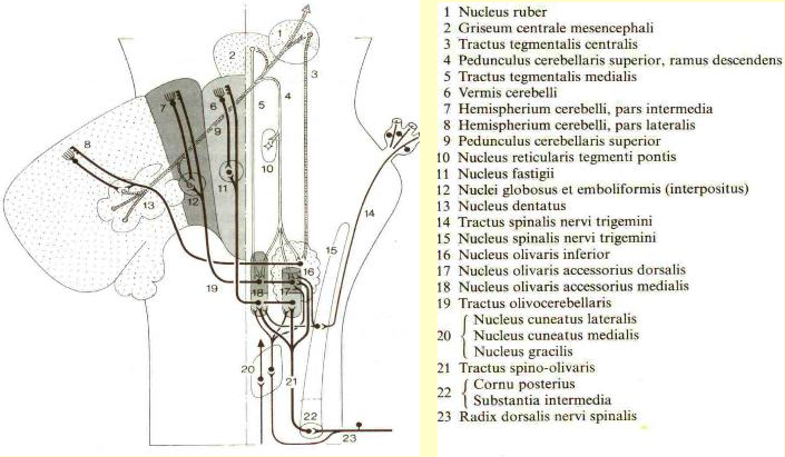
Hair cells activate primary projections of the vestibular system. Muscle spindles monitor parent muscle action and project to relay nuclei in the Central Nervous System (CNS).
T2E Primary neurons project from hair cells to vestibular nuclei as Idea Terms. Proprioceptive relay nuclei in the CNS also project as Idea Terms to the vestibular nuclei from all spinal levels.
T8E Parallel patterned response outputs that balance sensory vestibular inputs project to eye, neck, and spinal motor nuclei.
T5R The motor output to eye and neck muscles is reconciled with other inputs to these motor nuclei. The eyes may move to compensate for head movement or move separately. This output is regenerative since it is governed by connections established through prior learning of the individual and the species. The motor output to the neck and eyes is via the Medial Longitudinal Fasciculus (MLF). The motor output to the ventral motor horns of the spinal column is via the lateral vestibulospinal tract.
Cycle 2
T7R Memory of the sequence is stored in dendritic and synaptic protein synthesis of cells involved, as well as the Void. The synaptic connections evolve consistent with learning experience. At the same time related regenerative memories are spontaneously recalled in the reticular formation. This activates efferent projections to hair cells and muscle spindles.
T1R The regenerative efferent projections to hair cells and muscle spindles initiate a patterned motor simulation. In the projection to muscle spindles this takes place in gamma motor neurons that project to the spindles.
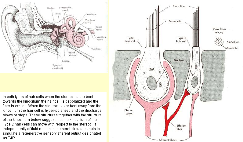
T4R An actual simulation takes place in hair cells and in neck and body muscle spindles. The hair cell simulation involves the active movement of the kinocilium in the Type 2 hair cell with respect to the current state of the Type 1 hair cells. The muscle spindle simulation likewise simulates a pattern of movement with respect to the current position of muscles.
T2R The simulations project as regenerative vestibular input and likewise corrective proprioceptive feedback to vestibular nuclei as a regenerative idea to restore balance.
Cycle 3
T8E 
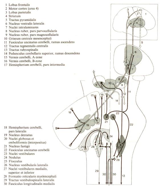
The simulations result in a synapse in the vestibular nuclei to a pattern of planned motor output to eye, neck and body muscles via the MLF and the lateral vestibulospinal tracts as in the T8E Term above. All Particular Set T8 Terms are expressive. (See Part 1).
T5E The output of vestibular nuclei to eye, neck and body spinal motor nuclei for corrective action is reconciled with other inputs to these nuclei as they project to muscles. This output is expressive since it is creatively planned by proprioceptive input. It is patterned action of the Host (1) that directs Organs (2) whereas in the regenerative sequence it is conditioned Organ (2) processes established through experience that direct the Host (1). (See Part 1)
T7E Memory of the action sequence is stored and expressive memories recalled similar to T7R above.
T1E There is a perception of the capacity to respond to ongoing vestibular needs. This involves the recovery of cells processes such as action potentials and the stabilization of fluid in semi-circular canals, preparatory for the next action sequence.

Keep in mind that muscle action is organized in the Central Nervous System (CNS). But sensory input, motor response in muscles, and related proprioceptive feedback at the spinal level are functions of the Peripheral Nervous System (PNS). In the context of spinal reflexes, the motor projection from the ventral cord is a T8E term and the T5 term follows in the muscular actions that produce proprioceptive feedback in the process.

In contrast, the CNS projection to the final motor centers such as the ventral horns of the cord is a T8E term in the CNS context and the T5 term follows as a the projection to muscles, not in the actions of muscles themselves as in the spinal reflexes. The projection to muscles is the final T5 leg of the CNS action sequence, whereas muscle action itself is the final T5 term leg in local action sequences, such as eye movements and spinal reflexes.

The arrangement allows for the integration of multiple motor inputs in the final motor nuclei in the CNS that project to muscles. If it was not this way, reconciliation and integration of conflicting motor inputs would not be possible. For example, conscious motor projections from the primary motor cortex of the cerebral hemispheres would not be able to override locally automated spinal responses in the cord that are not consciously determined.

As it happens, the cerebral motor projection to the ventral spinal motor area is a T8E motor term that is one sequence Step ahead of the spinal interneuron Idea Term T2 that also projects to the ventral motor horn. The cerebral T8E term can thus activate designated motor neurons in the ventral horn preferentially to spinal input, but it comes later. It takes one four Step Cycle to make a transition from one action sequence to another. The structure of the nervous system has evolved to function in accord with System 4 in just this way.

In conjunction with other inputs, body movement at all spinal levels is integrated via the lateral vestibular spinal tract in parallel with neck and head movements as shown above. There are three Particular Sets involved in the vestibular coordination of head, body, and eye movements. They integrate past and future as usual. The Universal Sets need not be considered in detail here since they work the same as at the spinal level illustrated in Part 1. It all meshes together very neatly consistent with our phenomenal experience.

B. Vestibular Integration with the Sensory and Motor Areas of the Cerebral Cortex

Vestibular projections to the cerebral cortex are still imperfectly known, however scattered cells in the medial, lateral, and superior vestibular nuclei project via a synapse in the thalamus to the primary sensory cortex of the cerebral hemispheres and to a second adjacent sensory association area behind it in the parietal lobe. While the projections in Section A above from the vestibular nuclei are patterned motor response projections that balance sensory inputs according to how T8E works, it has been suggested in some texts that these parallel projections to the sensory cortex should be considered sensory.

They are projections of motor action patterns regulating balance that must be consciously integrated with sensory input from all sources in working out an integrated planned System 4 Idea Term T2. This consciously planned Idea is topologically formulated in the primary sensory cortex. It projects in turn to the primary motor cortex where final motor instructions are topologically integrated in the T8E terms that project to the ventral horns of the spinal column where they synapse with the T5 terms that project out to the peripheral skeletal muscles.

In fact, the primary sensory cortex has motor characteristics, just as the primary motor cortex has sensory characteristics, as was emphasized by C.N. Woolsey, one of the early investigators of the motor and sensory areas. It is known from electrical simulation studies that there is a sensory core with a wider surround throughout the sensory cortex, and a motor core with a wider surround throughout the motor cortex. Motor responses are primary in motor areas and sensory responses in sensory areas, but each has characteristics of the other. (Published in Cerebral Localization and Organization, G. Shaltenbrand and C.N. Woolsey, Eds., University of Wisconsin Press, 1964.)

The vestibular motor characteristics that project to integrate head, body, and eye movements thus relay this information to the thalamus via the T8E term and on to the primary sensory cortex via the T5 term. The billions of microscopic nerve cells of the cerebral cortex are arranged in six layers with complex interconnections between them that are nevertheless suggestive of the six term sequence of the Particular Set transformations of Systems 4 and higher. It is these subsumed cortical processes that generate the coherent Idea plan T2 of the Sensory Cortex, taking account of all inputs.

The way System 4 works, T2 has a polar relationship with the resources of memory and recall, T7, which is the next System 4 Term following the T5 vestibular projection to the Primary Sensory Cortex. The recall process is obviously coupled to sensory input in a preceding sequence in order that we can subsequently respond appropriately to circumstance as it is presented to us. This requires that the T7 synapse is synchronous with the projection pattern of T4 synapses to the sensory cortex that results in the following T2 projection to the Primary Motor Cortex. Terms 8, 7, and 4 always occur together but in different Particular Sets. This has implications in the subsumed cortical processes where the vestibular T5 terms synapse. Idea development T2 is also tensionally coupled with the physical action term T5 in the active matrix of System 4 transformations. Terms 1, 2, and 5 always occur together in different Particular Sets.

Later, we will come back to the reciprocal relationships between the cerebral hemispheres and the cerebellum that intermesh with these vestibular inputs. For the present, it is relevant that the four vestibular nuclei play an analogous role to the four deep nuclei of the cerebellum.

C. Vestibular Projections to the Cerebellum

Vestibular inputs to the vestibular areas of the cerebellum follow two general routes as follows:

1. Via primary afferents from hair cells, by passing the vestibular nuclei, mossy fibers project directly to granule cells in the flocculo-nodular areas of the cerebellar cortex. Collateral branches project to the Fastigial cerebellar nucleus. This nucleus projects to the descending medial longitudinal motor tract via the reticular formation. It also projects via the thalamus to the premotor, primary motor, and parietal areas of the cerebral cortex as well as to the basil ganglia.

2. Primary afferents from hair cells project to the lateral, superior, medial, and inferior vestibular nuclei. They project in turn via mossy fibers to granular cells in the vestibular areas of the cerebellum, both directly and via a synapse in the inferior olive. The projections from the inferior olive are climbing fibers that synapse directly with Purkinje cells and these will be considered later.

C-1. Vestibular Projections Bypassing Vestibular Nuclei

Since it is known that the cerebellum is concerned with integrating vestibular function and conscious motor control with spinal proprioception, some parallels with spinal proprioception may be expected.

Proprioceptive sensory feedback from muscle spindles projects into the dorsal sensory horns of the spinal cord, but these neurons also send collateral branches to the ventral motor horns, thus bridging interneuronal connections within the spinal cord. System 4 indicates that the collateral projection to the motor horn designates a motor target pattern that is anticipated. (See Part 1) It readies these motor neurons to fire when and if the interneurons make the appropriate connections between sensory input from various sources and motor output.

They are also primed to fire depending on a compatible motor pattern from higher centers including the cerebral cortex. The degree to which the actual motor pattern that transmits to muscles matches the anticipated target pattern thus depends upon synchronous sequences of System 4. These are transmitted as Idea Terms T2 via interneurons in the cord to T8E motor neurons in the ventral cord, as well as by descending T8E motor patterns from higher brain centers. All these motor inputs must be mutually reconciled in the ventral cord.

The point here is that vestibular input from hair cells that bridges both the vestibular nuclei and the cerebellar cortex with collateral projections to the Fastigial Nucleus and vestibular nuclei can serve to prime a pattern of neurons in readiness to fire depending upon other Fastigial or vestibular inputs. Some of these inputs may come in System 4 Steps that precede or follow in synchronous sequences of System 4. We shall see below that there is an inverse pattern to the way this works in the cerebellum, since Purkinje cells are inhibitory. The Fastigial and vestibular nuclei play similar roles but with different patterns of projection.

Note: A very important point is that the cortex of the cerebelum cannot be considered as entertaining all System 4 Steps, since the only active output from it is inhibitory. It functions only at the Form level of the System 4 hierarchy. This is an ingenious invention since it facilitates the integration of inputs from many different sources. It does not say what to do. It selects from various inputs choosing what not to do. It either has no output at all from Purkinje cells, remaining neutral, or it has inhibitory output.

To simplify an otherwise complex description, it will be assumed that the direct projection of primary vestibular neurons to the cortex of the cerebellum is a regenerative sequence. This route by-passes the vestibular nuclei. It has a collateral branch to the Fastigial nucleus, and of course there is a parallel pathway that projects to the vestibular nuclei.

There are thus parallel pathways to the cerebellum and both pathways can have expressive and regenerative sequences. In the descriptions to follow, the Fastigial and vestibular nuclei may be interchangeable except that they project to different target patterns. The Fastigial nucleus will be used to illustrate the regenerative sequence and then the vestibular nuclei will be used in the expressive sequence. There will be more on this later to clarify the expressive and regenerative pathways.

With these points in mind, the System 4 sequence proceeds as follows with respect to the Fastigial nucleus. (Remember that there are three Particular Sets involved and they follow the same sequence, but starting in different Steps. In this Set, Step 1 starts with a T7R Term. See the Summary Chart near the beginning or Part 1.)

T7R - In Step 1, there is a pattern of regenerative recall T7R in reticular formation arousal.
T1R - T7R initiates a motor simulation T1R in efferent neurons projecting to hair cells in Step 2.
T4R - Type 2 hair cells simulate corrective action with respect to Type 1 hair cells, as R1 in T4R.
T2R - Primary vestibular neurons project as T2R in Step 4 directly to Cerebellar Granule cells with collaterals to the Fastigial Cerebellar nucleus.
T8E - The Fastigial nucleus reconciles inputs from other sources in motor projections ascending to the thalamus and descending to the reticular formation with a collateral projection back to vestibular nuclei. The Purkinje cells of the cerebellar cortex make inhibitory projections back to the Fastigial nucleus two Steps later. This integrates expressive and regenerative sequences from hair cells (since they alternate every other Step) along with other Fastigial inputs.
T5E - Motor projection proceeds from a synapse in the thalamus to motor and pre-motor areas of the cerebral cortex, as well as to the parietal lobe and the basal ganglia. Descending motor projections proceed from a synapse in the reticular formation to spinal levels via multi-synaptic tracts.
T7E - The vestibular and cerebellar sequence Steps are recorded in memory both in the Void and as protein synthesis that tends to modify the dendrite and axon connections of related cells accordingly, as an expressive sequence follows as outlined below.

Since the integrated pattern of motor output from the Cerebellar cortex to the Fastigial nucleus is only by inhibitory Purkinje cells, this output effectively carves away from the pattern of collateral T2E inputs from the hair cells two Steps later (assuming that both expressive and regenerative modes follow this pathway). The remaining T2R inputs, that are not inhibited by an earlier T2E projection, initiate the modified T8E outputs of the Fastigial nuclei to both descending spinal and ascending cerebral motor projections.

The ascending projection is by a one synapse pathway to arrive at cerebral motor centers as a T5E motor term. The descending projections are reticulated as a series of T8E motor projections until their final synapse on the motor horn cells of the spinal column. There they synapse with motor neurons that represent the final leg of T8E projection to the skeletal muscles. The descending T8E projections are a Step later in the System 4 sequence and tend to assume priority over the T2 spinal interneuron projections from the dorsal horn, but they arrive in later sequences accounting for a delay in rectifying balance. It should be noted that under normal circumstances that the influence of the vestibular regenerative mode can be relatively minimal compared to a gymnast or athlete who is well practiced.

Mossy Fiber Inputs:
(Adapted from Nieuwenhuys. Voogd. van Huijzen The Human Central Nervous System, Springer Verlag 1990)


C-2. Vestibular Projections via the Vestibular Nuclei

Consistent with the comments above, for the sake of simplicity, for the time being let us assume that the primary projections to the vestibular nuclei are an expressive mode of System 4 that alternates with the regenerative mode. If there is a parallel regenerative projection to the vestibular nuclei, it is easy enough to follow by simply substituting the vestibular nuclei for the Fastigial nucleus in the description above. We will come back to this later. As mentioned, the general pattern is analogous to the way the expressive and regenerative modes work via muscle spindles at the spinal level. Later, we shall see that there are complementary pathways via the inferior olive.

Allowing for alternating sequences in hair cells, as well as for one synapse in the vestibular nuclei, this means that there is an expressive sequence via the vestibular nuclei alternating with a regenerative sequence projecting directly to the cerebellum every two Steps. Since the projection via granule cells to inhibitory Purkinje feedback to the Fastigial nucleus takes two Steps, this facilitates the reconciliation of the expressive and regenerative modes. Purkinje inhibition from an expressive sequence modifies a regenerative pattern and vice versa. It moderates the transitions between modes into smooth patterns of motor projection. In the case of a parallel regenerative sequence via the vestibular nuclei, it works in an identical way except that the Purkinje projection is back to the vestibular nuclei rather than to the Fastigial nucleus.

The vestibular nuclei play a similar role to the Fastigial nucleus in this expressive sequence as compared with the regenerative sequence. There are various alternatives possible in some portions of the vestibular system as opposed to other portions, affecting some motor projections in different ways that others, although they all conform to the way System 4 works. This can direct experimental work to investigate the possible expressive and regenerative alternatives. For now, we will focus only on the expressive mode through the vestibular nuclei as follows:

T7E - Patterns of expressive recall T7E arouse in the reticular formation. (Step 3 of a Cycle)
T1E - Preparedness of cells to respond to needs by efferent projections to hair cells in Step 4.
T4E - Expressive sensory input from hair cells synapse with primary neurons in Step 1.
T8E - Motor outputs from the lateral nucleus project via the lateral vestibulospinal tract to the motor areas at all spinal levels. The superior, medial, and inferior nuclei project via the medial longitudinal fasciculus (MLF) to eye and neck motor nuclei. Motor outputs project to the inferior olive and also via mossy fibers to the cerebellar cortex. Both of these latter projections to the cerebellum result in a pattern of Purkinje inhibitory feedback two steps later. This reconciles expressive and regenerative motor projections from the vestibular nuclei, since they alternate every two System 4 Steps.
T5R - The motor projections T8E via the lateral vestibulospinal tract synapse in the spinal motor horns throughout the cord. As before, the T8E reticulated motor pattern projections coincide with T2 interneuron inputs to the motor cells in the ventral horns. These motor cells must reconcile these inputs from different sources that project as T8E Terms in the final spinal motor projection to the skeletal muscles. This is the same as with the descending cerebral motor projection T8E described for the regenerative sequence above, but it follows two Steps later. Direct Motor projections from the Primary Motor Cortex of the cerebral hemispheres also project to the ventral horns as T8E Terms. Both have priority over the T2 spinal interneuron projection from the dorsal horn as pointed out above. We may stumble when walking, but vestibular balance will tend to prevail over the next automated step and conscious intention quickly follows in a later sequence.
T7R - The vestibular and cerebellar sequence Steps are recorded in memory both in the Void and as protein synthesis in the dendrite and axon connections of related sequence cells, as a regenerative sequence follows as outlined above. Learning through experience elaborates on synaptic connections that reflect our remembered ability to perform in specific patterns.

The pattern of inhibitory output of Purkinje cells back to the vestibular nuclei is thus similar to the regenerative sequence where they project back to the Fastigial nucleus. The inhibitory Purkinje output likewise carves away from the T2E projections originating from hair cells and muscle spindles alike to the vestibular nuclei in the expressive sequence Steps just as it does in the regenerative sequence Steps to the Fastigial nucleus. Purkinje cells in part of the Vermis area of the cerebellum project back to the lateral vestibular nucleus that gives rise to the lateral vestibulospinal motor tract that projects as a T8E Term to all spinal levels. Purkinje cells in the nodulus and floccuus areas of the cerebellar cortex project back to the superior, medial, and inferior vestibular nuclei. Together, these nuclei project as T8E Terms to both the ascending and descending medial longitudinal tracts (MLF) as outlined in Section A above.

The Inferior Olive - Cerebellum Connections

Projections to the Inferior Olive

The inferior olive is a major structure in the brain stem that receives inputs from the following two main sources:

1. Collaterals of spinal somatic and proprioceptive sensory input from all levels. These all initiate in the PNS as T4 sensory input Terms of System 4 and project as T2 idea Terms to the inferior olive via a single synapse.

2. Collaterals from the superior, medial, and inferior vestibular nuclei arise as T4 sensory Terms from hair cells that project as T2 idea Terms to vestibular nuclei. They then project as T8E motor terms to the inferior olive.

Climbing Fiber Interconnections from Inferior Olive to Purkinje Cells and Deep Nuclei

The inferior olive projects via climbing fibers directly to Purkinje cells in nearly all areas of the cerebellar cortex with collateral projections to all four deep cerebellar nuclei. However, it projects preferentially from specific olive nuclei to the Vermis cortex, the Intermediate cortex, and the Cerebrocerebellar cortex. The respective collateral branches project to the Fastigial nucleus, the Globus and Emboliform nuclei, and the Dentate nucleus. Since Perkinje cells project back to these nuclei the cortex is bridged in a single Step as compared to two Steps via mossy fibers.

1. Spinal System 4 Steps via Inferior Olive

Sensory input from the spinal level comes once in each System 4 Step of each Cycle from one of the three Particular Sets. (See Chart above or Part 1). Sensory input from the head does not use the spinal nerves, but it follows analogous pathways. In Step 1 of each Cycle, it is always Somatic sensory input associated with touch, pain, temperature. Proprioceptive feedback from muscle spindles comes in Steps 2, 3, and 4 of each spinal Cycle and the nature of its origin is determined only by which Step it is in. For example, proprioceptive simulation of an action sequence always takes place in Step 3 of each Cycle. In Step 2, input from muscle spindles is generated by an expressive muscle action sequence, and in Step 4, by a regenerative muscle action sequence, but it still comes from the same muscle spindles. These latter two are also sensory inputs, and so far as higher centers are concerned, they are represented by T4 Terms, whereas at the spinal level, they are represented by a specific Relational Whole in the T5 Term providing direct sensory feedback that monitors muscle action in this spinal context. (See Part 1) Although the specific expressive or regenerative nature of the proprioceptive sensory inputs to the inferior olive may not seem very significant, in a later section we shall see that they are. In this section, it is very significant with respect to the Fastigial nucleus as pointed out below. With these obervations in mind, the sensory inputs to the inferior olive corresponds to the following System 4 sequence Steps:

T1 - Restoration of action potentials T1E occurs in Step 4 of each Cycle in readiness for a new sequence pattern at the beginning of the next Cycle. This is synchronous in both the spinal and vestibular pathways. In Step 2 of each Cycle, in one Particular Set there is a Gamma T1R motor simulation projecting to muscle spindles (See Part 1). In the same Step, there is also an efferent motor simulation projecting to hair cells. Both simulations arise as a pattern of spontaneous recall in the reticular system. We will look at the vestibular-olive projections in Section 2 below.
T4 - In Step 1 of each Cycle, primary somatic input from the PNS synapses in the CNS. In Steps 2, 3, and 4, proprioceptive sensory neurons project from muscle spindles in the PNS to a synapse in the CNS.
T2 - Collaterals of all secondary sensory neurons from the first CNS synapse project to the inferior olive in each Step of each Cycle. Note that secondary neurons from spinal levels also project via mossy fibers directly to the cerebellar cortex with collaterals to the deep nuclei in each Step, but one Step ahead of projections to the cerebellar cortex via the Olive route. This is very significant, since Purkinje inhibitory feedback is very specific and comes in one Step via the climbing fiber route and in two Steps via the mossy fiber route, which is more widely dispersed. This means that the respective patterns of Purkinje cell inhibition back to the deep nuclei are synchronous between mossy and climbing fibers. They arrive back at the deep nuclei together to simultaneously influence the patterns of projection from them.
T8E - The Inferior Olive projects to Purkinje cells in a highly specific manner with collateral projections to the deep cerebellar nuclei. Purkinje cells project back to the deep nuclei in one Step. They thus inhibit to carve a pattern of motor projections from the affected nuclei one Step later than collateral projections. Although mossy fiber projections get to the cortex one Step ahead of Inferior Olive projections, it takes two Steps to generate a more widely dispersed pattern of Purkinje cell inhibition back to the deep nuclei. The mossy fiber projections integrate more diverse areas of the body in more general ways.

Note that the collateral T8E projection to the Fastigial nucleus in Step 3 originates from an expressive somatic sensory input sequence T2E and it is synchronous with a direct T2R vestibular-Fastigial input from hair cells as outlined in C-1 above. They must find reconciliation in the pattern of projection from the Fastigial nucleus. This is essential because otherwise a conditioned pattern of expressive spinal responses would unilaterally prevail. This is strong evidence that the direct vestibular projection to the cerebellum that bypasses the vestibular nuclei is regenerative in this Step. The reverse is true in the following T8E Term two Steps later, because this spinal sequence originates in a pattern of regenerative muscle spindle simulations. This strongly indicates that the direct projection from vestibular hair cells is expressive in this Step. It also indicates that hair cell projections alternate between expressive and regenerative sequences in the pathway via the vestibular nuclei as well as via the pathway that bypasses them and projects directly to the Fastigial nucleus and cerebellum. This is very important.

T5 - All four deep nuclei project modified motor patterns ascending to motor, pre-motor, and parietal areas of the cerebral cortex as well as to basal ganglia via one synapse in the thalamus. The thalamus is the final ascending T5 terminal in all these sequences. Descending motor patterns project from the Fastigial nucleus to the reticular formation. It is reticulated via this multi-synaptic pathway to the motor areas of the entire spinal cord where it influences the final motor projection to skeletal muscles as before. This influence is delayed via the reticulation as compared with more direct routes via the vestibular nuclei. The reticulation can also influence the recall pattern of regenerative sequences in the next Step at various spinal levels.
T7 - The memory sequence is stored via protein synthesis elaborating on axon and dendrite interconnections between the cell processes involved in the whole sequence, as well as in the Void, as always. In the same Step, appropriate related memories are recalled. In the expressive sequence, the recall relates to the pattern of cells required in a conditioned response. In the regenerative sequence, it relates to the pattern of cells needed in a motor simulation. This alternating pattern as it projects up to the cerebral hemispheres will be governed largely by the return path from earlier input to the cerebral cortex via direct mossy fiber input to the cerebellum to be described below.
T1 - In the expressive mode, the restoration of the action potentials in the spinal pattern of nerve and muscle cells needed is reflected as an ability to respond. The response capacity may be affected by their exhaustion, in extreme cases, exhausting the whole person. In other cases, the response capacity may be hyper-sensitized to conditioned patterns of recall depending upon general circumstances, as in facing threatening or pleasing events. In the regenerative sequence, motor simulations are initiated from the reticular system. They project via efferent neurons to hair cells and also via gamma motor neurons to muscle spindles.

Climbing Fiber Interconnections via the Inferior Olive

(Adapted from Nieuwenhuys. Voogd. van Huijzen The Human Central Nervous System, Springer Verlag 1990)

2. Vestibular System 4 Steps via the Inferior Olive

Projections from the vestibular nuclei with expressive and regenerative sequences arrive in the inferior olive as T8E Terms in Steps 2 and 4 of each Cycle. For simplicity, we will deal with them together here. The same Terms project as collaterals to the Fastigial nucleus which is closely associated with the vestibular system.

T1 - In Step 2 of each Cycle, there is a T1R motor simulation projected to the hair cells that initiates a patterned simulation in the hair cells. In Step 4 of each Cycle, the T1E pattern of efferent projections to hair cells readies the next action sequence.
T4 - In Step 1 of each Cycle, the hair cells respond in the expressive sequence. Motion of the head produces a pattern of sensory input T4E to the vestibular nuclei that makes conditioned synapses there. In Step 3, the Type 2 hair cells simulate a corrective T4R resposne with respect to the Type 1 hair cells. The resulting pattern of synapses is not automatically conditioned, but anticipates a better response pattern.
T2 - In Step 2, a conditioned pattern of response is projected to the vestibular nuclei as an expressive idea T2E. In Step 4, the T2R projection is a regenerative idea.
T8E - In the Particular Sequences, the T8 Terms are always expressive since they all result in motor patterns. (Only the Primary Universal Set has a T8R Term that distributes resources according to a priority of needs. See Part 1) Projected motor patterns have either an expressive or a regenerative history hower. They project to the inferior olive with a collateral projection to the Fastigial nucleus. We saw in the section above how the direct route bypassing the vestibular nuclei to the Fastigial nucleus synchronized in Steps 1 and 3 as expressive and regenerative inputs with alternate T2R and T2E projections from the spinal levels. Now, with one synapse in the vestibular nuclei, we find that this T8E projection has alternate expressive and regenerative histories. This complements proprioceptive sensory feedback to the Fastigial nucleus from muscle action terms having alternate expressive and regenerative histories and that input the Fastigial nucleus as T2R and T2E Terms respectively in Steps 2 and 4. Again, we have a situation where expressive and regenerative modes must be mutually reconciled to result in smooth performance.
T5 - From the synapse in the inferior olive, there is a climbing fiber projection directly to the Purkinje cells in the vestibular areas of the cerebellar cortex. This is not a progressive sequential System 4 Step however since there is either no feedback from the Purkinje cell or inhibitory feedback, as mentioned previously. This function level feedback may be either to the Fastigial nucleus or to the vestibular nuclei depending on inputs. As feedback to the lateral vestibular nucleus it influences a modified pattern of motor projection that must find reconciliation with the T2 spinal inputs and T2 hair cell inputs to that nucleus that result in the T8E projections to the spinal nuclei via the lateral vestibular-spinal tract. This over-riding influence comes in a later sequence of projections than the initial inputs to the lateral nucleus however. Nevertheless, the expressive or regenerative history of the T5 Term will require reconciliation with the alternate T2R and T2E Terms that project to the lateral nucleus from spinal muscle spindles as well as from hair cells. Terms 1, 2, and 5 always occur together and T2 and T5 are always tensionally coupled.
T7 - As usual, the System 4 expressive and regenerative sequences contribute synaptic interconnections via protein synthesis in nervous system organs and muscles linked with and keyed to memory storage in the Void. A new associated pattern of recall will be initiated via reticular system arousal. The pattern will be appropriate to new sensory input since Terms 8, 7, and 4 always occur together and T7 and T4 are tensionally coupled.

Cerebellum Outputs

(Adapted from Nieuwenhuys. Voogd. van Huijzen The Human Central Nervous System, Springer Verlag 1990)

Cerebral Pathways to and from the Cerebellum

Return Pathways to the Cerebellum

The largest contingent of mossy fiber projections to the cerebellum comes from the Pontine nuclei which receive somato-topically organized projections from the whole Cerebral Cortex. The densest projections come from the following areas: the pre-motor area, the primary and secondary motor areas, the primary and secondary sensory areas, and the sensory association areas. The pre-motor area integrates input to the topological motor areas, in a similar way that the sensory association area integrates input to the topological sensory areas.

They project to most areas of the cerebellum with the possible exception of the nodulus and they send collateral fibers to the deep nuclei, especially the dentate nucleus. The somato-topical projections overlap with those from spinal levels in the homunculi, which extend into the lateral hemispheres of the cerebro-cerebellum. The dentate, globose, and emboliform nuclei project back via the thalamus to the cerebral cortex in a similar pattern to which they arose. The number of term transformations and related synapses involved in the return journey correspond to six sequential Steps of System 4. The return feedback is thus positioned to influence integrated motor outputs from the cerebral cortex one sequence later. Again, this means that the expressive and regenerative modes must find reconciliation.

Cerebral Sensory Return Pathway

For example, output from the Primary Sensory Cortex represents either an expressive or regenerative T2 Idea term following the assimilation of T4 sensory input to it from many sources via the parietal lobe. It projects to synapse with the Primary Motor Cortex resulting in a descending motor pattern T8E. The parallel projection to the Pontine nuclei also results in a pattern of T8E projection to the deep cerebellar nuclei with a collateral projection to the granule cells of its cortex. Two Steps later, a pattern of Purkinje inhibition projects back from the cortex to the deep nuclei which now have received the next T8E projection from the Primary Sensory Cortex. This pattern of inhibition modifies the latter input to the deep nuclei which project back as alternating T5R and T5E Terms to the thalamus.

The two Step inhibitory pathway means that alternating expressive and regenerative sequences must be reconciled in the deep nuclei. The T5 projection to the thalamus results in a T7 Term, with the sequence stored as protein synthesis as well as in the Void as described before. The pattern of recall at this point will be tensionally coupled to the assimilation of sensory input T4 in the parietal sensory association area which supplies the Primary Sensory Cortex. This pattern of spontaneous recall results in T1 projection from the thalamus back to the Primary Sensory Cortex. Expressive and regenerative modes of T1 will alternate in this projection to synapse as either T2E or T2R Terms back with the alternate mode of the T2 Term currently being formulated in the Primary Sensory Cortex. In other words, the regenerative and expressive modes find mutual reconciliation spanning one complete six Step sequence. The anticipated idea pattern of regenerative action is thus reconciled smoothly with the conditioned expressive idea pattern of action. Emotional input via the limbic system works in synchronous accord.

Cerebral Motor Return Pathway

We can follow input from the Primary Motor Cortex in the same way. It generates a T8E Term with either an expressive or regenerative history which projects to the motor areas of the ventral spinal cord. The parallel projection to the Pontine nuclei is also a T8E Term. This results, after the synapse, in a T5 Term projection to the deep cerebellar nuclei and to the granule cells of its cortex. With the synapse in the deep nuclei, the motor sequence is recorded in memory as above, along with the spontaneous recall, T7, of a new pattern that is also tensionally coupled to the T4 sensory association area of the parietal lobe. (The latter has intimate dense projections to the pre-motor area, which in turn, projects as a T2 Term to the Primary Motor Cortex resulting in the initial T8E projections.) The pattern of recall in the deep nuclei is thus very relevant to the assimilation of idea in the Primary Sensory Cortex. It activates a pattern of T1 projection back to the thalamus. This can be either expressive, preparing a conditioned pattern of related cells, or a regenerative simulation that more creatively designates an original pattern. However, the two Step pattern of Purkinje cell inhibition requires that the expressive and regenerative sequences again be reconciled in the deep nuclei. The modified T1 pattern of projection to the thalamus alternates between expressive and regenerative modes and it results in corresponding T2E and T2R alternating projections back to the Primary Motor Cortex where they synapse to produce descending T8E motor Terms to the spinal ventral horns, as well as another parallel sequence back to the cerebellum. In this way, the return input from the cerebellum to the Primary Motor Cortex complements the reconciled input from the Primary Sensory Cortex and alternating T2E and T2R inputs to the Primary Motor Cortex.

Feed Forward and Feed Back Cerebellar Cortical Processes

The projections from the Primary Sensory Cortex come to the deep nuclei one sequence Step earlier and later than those from the Primary Motor Nuclei, and vice versa. So, the deep nuclei are only involved with one or the other at the same time. In examining the cell structure of the cerebellar cortex above, we have seen that there are feed back and feed foward processes at work within the cerebellar cortex. These processes can thus serve to facilitate the sensory assimilation of an integrated idea with the motor execution of the plan.

This has synchronous implications with inputs from other sources. For example, spinal sensory input T4 arrives after one CNS synapse as a T2 Term at the deep nuclei, which results in a T8 motor projection back to spinal levels. But projections from the Primary Sensory cortex to the deep nuclei arrive as T8 Terms, that is, one System 4 Step later than the T2 Term input from spinal levels. The feed back and/or feed foward synaptic processes within the cortex can thus serve to reconcile spinal sensory inputs with those that have been integrated from prior sequences in the Cerebral Hemispheres. This further facilitates smooth transitions from one action sequence to the next, spanning sequentially spatial orientation and increments of time.

Bilateral Integration

Cortical processes in the cerebellum can also facilitate the smooth bilateral integration of motor patterns as they relate to the two sides of the body. This works in conjunction with interneuron connections between the vestibular nuclei on the left and right sides of the body and likewise with the deep cerebellar nuclei on one side as opposed to the other. Inputs to the cerebellum can be bilateral, contralateral, or ipsilateral depending upon their source. But in general, the motor pattern on one side generally acts as a referent to the motor pattern on the other just as one foot step takes the other foot as a point of departure in walking. The feedback and/or feed foward cortical processes may be employed in this bi-lateral integration of behavior.

Once one is familar with the correspondence between System 4 and the way the nervous system has been structured to function synapse by synapse, anyone with the patience can explore these and other pathways at their leisure. It nevertheless demands a lot of concentration.

Summary

The cerebral hemispheres invest us with conscious experience. It invests us with conscious knowledge of our intuitive, rational, and emotional experience. This general triadic organization corresponds with the right hemisphere of the neo-cortex, the left hemisphere of the neo-cortex, and the ancient limbic cortex respectively.

There are commissures that connect the hemispheres of the neo-cortex and separate commissures that connect the two hemispheres of the limbic cortex. (See Inside our Three Brains) Within each hemisphere there are both primary and secondary sensory and motor areas that accomodate a self-similar arrangement of conscious intuitive, rational, and emotional experience within each hemisphere, but subsumed within the over-riding context each of these three focal points as they apply to the whole cortex.

For example, we can have socially oriented intuitions confined to the left brain that relate to rationalizations employing language and that may result in explicit behavior. We can also seek holistic right brain intuitive insight into the workings of the cerebral hemispheres that can find left brain expression as science. The former is confined to the left brain, while the latter involves the integrated workings of the whole cortex. The main point here is that the cerebral cortex is an instrument that allows us to consciously simulate experience in thought. This allows us to span and integrate space-time events from practicing our golf swing to understanding our own historical evolution. There has traditionally been a strong emphasis on dominant left brain social thinking as opposed to right brain intuitive insight. The System however provides a paradigm as to how we can relate more constructively to our social, spiritual, and natural environments.

As we have seen, the cerebellum reconciles cerebral processes with spinal and vestibular inputs, depending upon our level of physical activity. There are also other primary sensory inputs to the process, including vision, hearing, taste, and smell. System 4 can be applied to these sensory systems piecemeal synapse by synapse, although subsumed higher systems in the cerebral cortex are also involved, such as in generating virtual images in the vision process.

The autonomic nervous system also has sensory and motor neurons and System 4 can be applied to it piecemeal as well. Its integration with the somatic nervous system involved System 5 at the limbic level, however. System 5 has twenty terms and is considerably more complex in the way it works as an elaboration of System 4. Virtual images, whether visual, conceptual, intuitive, or emotional, first appear in System 5.

It should be mentioned that there are other variants to System 4 that we have not explored. The expressive and regenerative variants involve Centers 1 and 2 changing places in the Particular Terms. There are expressive and regenerative involutionary variants also where Centers 3 and 4 change places in the Particular Terms which constitute an involutionary variant that is inconsistent with the evolutionary variant discussed here. Expressive and regenerative modes are reversed. Values become inverted, such that there is a bi-polar moral dilemma at the roots of perception. Values become inverted.

Processes of disease and decay are related to involutionary variants, but they are essential to metabolism also. Other varients are possible but they have no conscious meaning. They can be related to sleep or dream states.

The above should be sufficient to demonstrate the System as a new paradigm that can prove of great value in its practical application to science.